
Chronic spontaneous urticaria (CSU) are kind of like your least favorite aunt. They show up unannounced and stay well past their welcome. These red, itchy welts have no obvious cause and last for 6 weeks or longer. And the effects of the continuous itching caused by CSU go far beyond just your skin. Living with CSU can affect your mental health, as well as your work and personal life. Symptoms of chronic spontaneous urticaria CSU isn’t predictable. Unlike hives that appear when you’ve been exposed to something you’re sensitive or allergic to, CSU hives come and go, seemingly at random. And they can stay for a long time. This means you might be having a “good” week, with few or no hives and you make plans to go out. Suddenly the hives appear, making you miserable. Or they could come out just before a big presentation at work or a much anticipated (and needed) vacation. This unpredictability can make you uncertain about making plans and frustrate you if you need to cancel them. It also can affect your relationships if those around you don’t understand how hard it can be to have recurring hives like this. Here are four ways living with CSU might affect your life. Increased anxiety and depression Researchers have found that people living with CSU have higher rates of anxiety and depression — up to six times higher — than those who don’t have the condition. If left untreated, depression can lead to physical problems, including heart disease and stroke. Both anxiety and depression can also affect your ability to get medical help, follow treatment plans, go to work or school, and take care of yourself and your family. A form of talk therapy, cognitive behavior therapy (CBT), is helpful for many people with anxiety and depression. The goal with this type of therapy is to find coping strategies and help you manage your thoughts and feelings. Speak with your healthcare provider (HCP) about getting help if you’re experiencing anxiety or depression. There are also groups that can offer support, such as WeCU and the Allergy & Asthma Network. Sleep deprivation We know that if we have pain, our sleep can suffer, but many people don’t realize how disruptive itching can be, so they might not understand how tired or fatigued you are. More than half of people with CSU can’t sleep properly. This in turn can worsen depression and anxiety, as well as other mental health issues. Sleep deprivation can also increase your risk of having accidents, injuries and long-term health issues, like heart disease and some types of cancer. If CSU is affecting your sleep, it’s important you speak to your HCP about it. If you’re taking second-generation antihistamines, which don’t make people sleepy, your HCP might suggest adding another one at night that does cause sleepiness. There may be other medication options that can help, too. Working on mind-body and relaxation approaches might be helpful. There are many self-help options to help people sleep, but working with a therapist might be the best way to start, especially one who works with people living with CSU or similar health problems. Reduced enjoyment of social and work life There are several reasons CSU might have a strong impact on your social life, work or education. Adults with CSU miss more work than those with other allergy-related conditions, and they don’t perform as well while they’re at work. According to one study, those with mild CSU lost about 12% of their work productivity, and those with severe CSU lost as much as 44%. The same happens with children with CSU. They do worse at school than their classmates who don’t have these long-term hives. Daily tasks and intimacy can be affected as well. It’s not unusual for some people with CSU to have trouble keeping up with personal care, family life and housekeeping. Sexual activity and intimate relationships can also become difficult. Maintaining a relationship can be challenging if you’re self-conscious of how your skin looks, you’re itching all the time, you’re fatigued, and your mental health is affected. Hobbies and just getting out to have fun are usually a good way to relax and recharge. But if you’re living with CSU, they might be the last thing you’re thinking of. Finding clothing that feels comfortable can be tough. Tight clothes rub against your skin and some clothing is made of irritating fabrics, like wool, or they have textures or seams that can feel uncomfortable on your skin. If you have a particular style that you like to follow, it can be disappointing if you have to choose looser fitting clothing in different fabrics that don’t match what you’d like to wear. If you have to wear a uniform at work or school, this can also be an even bigger problem, and you might need to ask for accommodations either in style or fabric. Increased risk for infection Although it’s not common, if you scratch your hives enough to break the skin, you can get an infection, which can become serious if not treated. If you do break the skin, be sure to clean the area, apply antibiotic ointment and cover the broken skin to protect it. See your HCP if you develop any redness or swelling, or if you see any discharge or pus coming from the area. Taking charge of CSU CSU is more than “just hives.” It has a significant impact on your life. Discussing your condition with your HCP to ensure that you find the right medication to control your hives and seeking support from the people around you as well as groups and communities that understand CSU can go a long way in helping you live a full life with this chronic condition. This educational resource was created with support from Regeneron and Sanofi. From Your Site Articles Related Articles Around the Web Source link

Nominations open for Healio’s Disruptive Innovators in GI
Add topic to email alerts Receive an email when new articles are posted on Please provide your email address to receive an email when new articles are posted on . “ data-action=”subscribe”> Subscribe We were unable to process your request. Please try again later. If you continue to have this issue please contact customerservice@slackinc.com. Back to Healio Healio’s ninth annual Disruptive Innovators Awards for gastroenterology and hepatology will be presented this fall, honoring the pioneers whose groundbreaking work and bold ideas have redefined the field. Awardees will be recognized during a prestigious awards ceremony at the ACG Annual Meeting, scheduled for October 9-14 in Nashville. Healio Gastroenterology and its collaborators unveiled its 2025 class of Disruptive Innovators Award recipients during the ACG Annual Meeting in Phoenix. Credit: Erin T. Walsh, MA The event will be hosted by Healio Chief Medical Editor Edward V. Loftus Jr., MD, and Ugo Iroku, MD, MHS, a co-founder of the Association for Black Gastroenterologists and Hepatologists. Healio is now accepting nominations in nine categories: Lifetime Disruptor, presented to a physician who consistently pushed the field forward through innovative treatments, practice management, patient care or research; Rising Disruptor, which recognizes an up-and-coming physician who is disrupting the status quo in the field through new techniques, new thoughts, questioning methods or breakthrough research; Clinical Innovation, presented to a physician or institution that changed the face of gastroenterology practice, providing an example of how patient care can be bettered through changes in administration, technique or delivery of value-based care; Woman Disruptor of the Year, presented to a woman in the field who has emerged as a leader and example to younger women of how a successful career can unfold; Social Media Influencer, which recognizes a health care professional who has made a positive impact on social media, become a trusted resource for his or her peers, and led the tidal change in health care provider use within gastroenterology; Healio Patient Voice, presented to a patient advocate or advocacy group that moved the needle with regard to discussion in the public sphere, better communication between patients and providers, and/or advocacy for legislative or regulatory action; Health Equity Award, which recognizes a physician who has made meaningful changes to overcome the social determinants of health in gastroenterology; The Partner in Practice Award, presented to an allied health provider — dietitian, gastropsychologist, nutritionist, nurse practitioner or physician assistant — whose innovative approaches improve patient outcomes or quality of care; and Industry Breakthrough, presented to a product that stands out as a major disruption to the practice of gastroenterology. Would you like to nominate a mover, shaker or industry name-maker who has spurred innovation or disrupted the practice of medicine to improve patient health or improve quality of care? Email nominations to Editorial Director Robert Stott at rstott@healio.com. Please include the nominee’s name and institution, along with the award category for which you believe the nominee is most deserving. Published by: Ask a clinical question and tap into Healio AI’s knowledge base. PubMed, enrolling/recruiting trials, guidelines Clinical Guidance, Healio CME, FDA news Healio’s exclusive daily news coverage of clinical data Learn more Add topic to email alerts Receive an email when new articles are posted on Please provide your email address to receive an email when new articles are posted on . “ data-action=”subscribe”> Subscribe We were unable to process your request. Please try again later. If you continue to have this issue please contact customerservice@slackinc.com. Back to Healio Source link
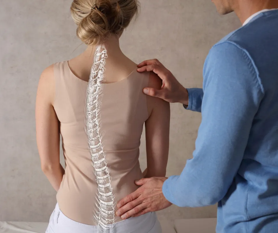
Interlaminar Spinal Fixation Systems
If you suffer from chronic back pain or have a spinal injury, you know firsthand how debilitating it can be. Fortunately, advances in medical technology have led to a range of treatments that can alleviate pain and restore function to the affected area. Interlaminar Spinal Fixation Systems are medical devices composed of two interbody spacers and a linking rod made of titanium. They helps reduce pain, prevent further damage, and promote spinal fusion. Furthermore, they are minimally invasive and have been shown to be effective in clinical studies. Join us as we take a closer look at the interlaminar spinal fixation system and how it can help improve outcomes for those with spinal instability or degenerative disc disease. What is an Interlaminar Spinal Fixation System, And What Are Its Components? Interlaminar Spinal Fixation Systems are spinal stabilization systems designed to promote spinal fusion and provide stability to the lumbar spine. The system consists of two interbody spacers and a linking rod made of titanium, which are implanted through a minimally invasive procedure. The interbody spacers are placed between two adjacent vertebrae to restore the natural spacing and alignment of the spine, while the linking rod connects the two spacers to stabilize the spine. The system’s design allows for controlled movement while the spine heals and promotes the growth of new bone tissue. These devices aim to reduce pain, prevent further damage, and improve spinal stability, leading to better outcomes for patients with spinal instability or degenerative disc disease. It’s also an effective alternative to traditional spinal fusion surgery, with fewer complications and faster recovery times. What are the Benefits of Interlaminar Spinal Fixation Systems? This system offers several benefits for patients with spinal instability or degenerative disc disease. Stabilization of the Spine: The first and most significant benefit is the stabilization of the spine, which can reduce pain and prevent further damage. The device does this by attaching itself to vertebrae and not allowing them to move, thereby reducing instances of further spinal damage, pain, or even inconvenience. Promotion of Spinal Fusion: Additionally, the system promotes spinal fusion, which is the natural process of two adjacent vertebrae fusing together. The interbody spacers used create the optimal environment for new bone tissue growth, leading to a more stable spine over time. Minimally Invasive Procedure Another significant benefit of spinal fixation systems is that it is a minimally invasive procedure. Unlike traditional spinal fusion surgery, which requires larger incisions and longer recovery times, This procedure can be performed through a smaller incision. This means that patients experience less pain, have shorter hospital stays, and can return to normal activities faster. Overall, spinal fixation systems offer a safer, more effective alternative to traditional spinal fusion surgery, providing patients with long-lasting relief from spinal instability, chronic spinal pain, and degenerative disc disease. Additional benefits include the utilization of local anesthesia, conservation of bone and soft tissue, decreased likelihood of epidural scarring and leakage of cerebrospinal fluid, a shorter hospitalization and rehabilitation duration, and the possibility of reversing the surgical procedure without impeding future surgical alternatives. Types of Spinal Fixation Systems The most popular system is StabiLink. It is an innovation by Southern Spine, a celebrated global manufacturer of spinal and thoracic inserts. Their innovations include the following: StabiLink® MIS Interlaminar Spinal Fixation System This implant is placed away from the neural and other elements of the spinal cord. The StabiLink® MIS Interlaminar Spinal Fixation System boasts several design features that enhance its performance and effectiveness. The implant has a small diameter with a wide-spike design, containing 16 spikes per implant, which spreads the load over a larger area. This feature increases the implant’s load-sharing capacity during both static and fatigue testing. The Laminar Lock Design is another notable feature that limits movement in all three planes, including lateral bending, axial rotation, and flexion/extension. It offers a wide range of implant designs and sizes for optimal anatomical fit. The low profile of the implant also enables access to facet joints and other surrounding anatomy. The torque-controlled locking mechanism results in secure fixation of the implant. Overall, StabiLink’s design features ensure greater stability, accuracy, and long-term effectiveness for patients undergoing spinal stabilization surgery. How Are Interlaminar Spinal Fixation Systems Inserted? During the procedure, a small incision measuring 2–4 cm is made using a precision guided inserter/compressor. This instrument is designed to streamline the implant placement process, making it more straightforward and accurate for surgeons. The precision guided inserter/compressor is an all-in-one instrument that eliminates the need for multiple instruments, including bulky compressors. It allows for the implant to be safely placed with or without the removal of the interspinous ligaments. As a result, the overall procedure time is reduced, and the implant insertion and compression are achieved with greater ease and accuracy. The Risks Involved As with any procedure, Interlaminar Spinal Fixation Systems come with their own risks. Risks may include wound infection, post-surgical CSF leak, blood clots, etc. It is important to note that these instances are rare, and, have a far lower probability of occurrence when compared to traditional spinal decompression surgeries. If you or a loved one suffers from degenerative disc disease or chronic back pain, we recommend visiting Pain Treatment Centers of America at one of our many locations. Our medical and ambulatory surgical expertise has helped thousands of patients, and we would be delighted to help you determine the best option for you. For more information, call us at (844) 215-0731 today! Source link

WomenTalk: Matters of the Heart
This episode is all about heart health — and why it’s something women can’t afford to ignore. Heart disease is the number one killer of women, yet so many of us don’t realize our risk or know what to look for. We’re joined by Martha Gulati, M.D., a leading expert in preventive cardiology and women’s heart health. She’s the director of the Women’s Heart Center at Houston Methodist and a nationally recognized voice in helping women better understand and protect their heart health. This episode was produced with support from Amarin. Watch more WomenTalk episodes >> Source link

FDA approves Hepcludex, first treatment for chronic hepatitis D
Add topic to email alerts Receive an email when new articles are posted on Please provide your email address to receive an email when new articles are posted on . “ data-action=”subscribe”> Subscribe We were unable to process your request. Please try again later. If you continue to have this issue please contact customerservice@slackinc.com. Back to Healio Key takeaways: Hepcludex injection was approved for chronic hepatitis D without cirrhosis or with compensated cirrhosis. It is the first FDA-approved treatment for the disease. Editor’s note: This is a developing news story. Please check back soon for updates. The FDA approved Hepcludex injection for the treatment of chronic hepatitis D infection in adults without cirrhosis or with compensated cirrhosis, according to a press release. The FDA approved Hepcludex injection for the treatment of chronic hepatitis D. Hepcludex (bulevirtide-gmod, Gilead Sciences) is the first FDA-approved treatment for the condition. “Today’s approval fills a critical gap in care for patients with chronic HDV infection, who until now have had no FDA-approved therapies available,” Wendy Carter, DO, acting director of the Office of Infectious Diseases at FDA’s Center for Drug Evaluation and Research, said in the release. “For individuals living with this chronic viral infection, this new treatment option offers hope in managing a disease that can rapidly progress to serious liver complications.” The approval was based on data from the phase 3 MYR301 trial, which tested once-daily Hepcludex 8.5 mg. At 48 weeks, 48% of patients given Hepcludex met the primary efficacy endpoint of combined response vs. 2% of those who underwent a 48-week treatment delay. The drug comes with possible side effects including anaphylaxis, injection site reactions, headaches, abdominal pain, fatigue and itching. There is a boxed warning that discontinuing the drug can severely, acutely exacerbate HDV and hepatitis B virus infection. The FDA previously granted Hepcludex breakthrough therapy designation and orphan drug designation. Published by: Ask a clinical question and tap into Healio AI’s knowledge base. PubMed, enrolling/recruiting trials, guidelines Clinical Guidance, Healio CME, FDA news Healio’s exclusive daily news coverage of clinical data Learn more Add topic to email alerts Receive an email when new articles are posted on Please provide your email address to receive an email when new articles are posted on . “ data-action=”subscribe”> Subscribe We were unable to process your request. Please try again later. If you continue to have this issue please contact customerservice@slackinc.com. Back to Healio Source link

What Is Lumbar Radiculopathy?
Have you ever experienced shooting pain in your lower back or legs that seems to come out of nowhere? If so, you may be suffering from lumbar radiculopathy. This common condition affects many people and can cause a range of symptoms, from mild discomfort to severe pain and immobility. In this article, we’ll explore what lumbar radiculopathy is, what causes it, and how you can manage your symptoms to regain your mobility and improve your quality of life. What Is Lumbar Radiculopathy? Lumbar radiculopathy, also known as sciatica or neurogenic sciatica, is a condition that results from compression or irritation of one or more of the nerve roots in the lower back. These nerve roots exit the spine and travel down the legs, and when they become irritated or compressed, they can cause pain, numbness, tingling, and weakness in the lower back, buttocks, and legs. It is a common condition, affecting millions of people worldwide, and can significantly impact a person’s quality of life if left untreated. What Are the Causes of Lumbar Radiculopathy? Lumbar radiculopathy is typically caused by compression or irritation of the nerve roots in the lower back, although the underlying causes can vary. Some of the most common causes of lumbar radiculopathy include the following: Herniated disc: A herniated disc occurs when the soft center of the spinal disc bulges or ruptures, putting pressure on the nearby nerve roots. Spinal stenosis: Spinal stenosis is a narrowing of the spinal canal, which can put pressure on the spinal cord and nerve roots. Degenerative disc disease: This condition occurs when the discs between the vertebrae in the spine begin to deteriorate, causing them to lose their cushioning ability and putting pressure on the nerve roots. Spondylolisthesis: Spondylolisthesis is a condition where one vertebra in the spine slips out of place, putting pressure on the nearby nerve roots. Trauma or injury: Trauma or injury to the lower back can cause lumbar radiculopathy, especially if it results in a herniated disc or other spinal damage. Tumors or infections: Rarely, tumors or infections can cause compression or irritation of the nerve roots in the lower back, leading to lumbar radiculopathy. It’s important to note that lumbar radiculopathy can also be caused by lifestyle factors such as poor posture, lack of exercise, or obesity, which can put excess strain on the lower back and contribute to the development of the condition. What Are the Symptoms of Lumbar Radiculopathy? Lumbar radiculopathy can cause a range of symptoms, which can vary depending on the location and severity of the nerve root compression or irritation. Some of the most common symptoms of lumbar radiculopathy include (but are not limited to) pain, numbness or tingling, weakness, loss of reflexes, or difficulty with bowel or bladder function in rare cases. It’s important to note that the symptoms of lumbar radiculopathy can come and go, and may be aggravated by certain activities or positions, such as sitting or standing for long periods of time. If you’re experiencing any of these symptoms, it’s important to see a doctor for a proper diagnosis and treatment. What Are the Treatments for Lumbar Radiculopathy? Lumbar radiculopathy can cause significant pain and discomfort in the lower back, and it’s important to seek treatment in order to manage the symptoms and prevent further damage. There are many different treatment options available for lumbar radiculopathy; read on for a few of the more common treatments: Physical Therapy Physical therapy is a common treatment for lumbar radiculopathy, as it can help to reduce pain and improve mobility in the affected area. Physical therapy for lumbar radiculopathy may involve a variety of techniques, including: Exercises: Specific exercises may be prescribed to help stretch and strengthen the muscles in the lower back and legs. These exercises can help to improve range of motion, reduce pain, and improve overall function. Manual therapy: This technique involves hands-on manipulation of the muscles and joints in the affected area. This can help to reduce pain and improve flexibility. Modalities: Various modalities such as ice, heat, ultrasound, and electrical stimulation may be used to help reduce inflammation and pain. Education: Education on proper body mechanics, postures, and ergonomics is an important component of physical therapy. The goal of physical therapy is to help improve function and reduce pain in the affected area, which can improve the quality of life of people with lumbar radiculopathy. A physical therapist will work with the patient to develop an individualized treatment plan that takes into account their specific needs and goals. Drug Therapy Drug therapy is a common treatment for lumbar radiculopathy, which involves the use of medications to relieve pain, reduce inflammation and improve overall function. The medications used in drug therapy can be prescription or over-the-counter (OTC) and can include the following: Non-steroidal anti-inflammatory drugs (NSAIDs): These medications, such as ibuprofen and naproxen, can help to reduce inflammation and relieve pain. Muscle relaxants: These medications can help to reduce muscle spasms and improve mobility in the affected area. Opioids: In severe cases, opioids may be prescribed for short-term pain relief. However, due to the risk of addiction and other side effects, opioids are generally not the first line of treatment. Antidepressants: Some antidepressant medications, such as tricyclic antidepressants and duloxetine, can help to relieve chronic pain. Anti-seizure medications: Medications such as gabapentin and pregabalin can help to relieve nerve pain associated with lumbar radiculopathy. It’s important to note that while drug therapy can be effective in relieving pain and inflammation, it may not be a long-term solution. Injection-Based Treatment Injection-based treatments are often used to treat lumbar radiculopathy when conservative measures such as physical therapy and medication do not provide adequate relief. The injections may contain anti-inflammatory medications, such as corticosteroids, which are injected directly into the affected area to help reduce inflammation and pain. Other types of injections may be used to help block the pain signals from the affected nerve. Living with lumbar radiculopathy can be challenging, but it doesn’t have to

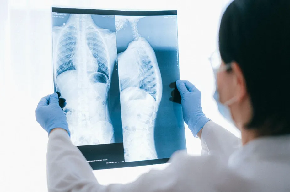
Nonsmoker Finding Lung Cancer Early
As told to Erica Rimlinger At age 43, I had very few risks for lung cancer, if any. I ran regularly, ate well and had never puffed a cigarette. I’d also never been exposed to secondhand smoke or dangerous levels of air pollution. In fact, my dad was a lung doctor who warned me since early childhood about the dangers of smoking and all the other potential threats to my lung health. So imagine my surprise when, after a quick run, I arrived for a CT scan that was sure to be routine and walked away with a diagnosis of stage 1 lung cancer. Just one hour before, I’d been running my usual route, with no airway problems at all. I wasn’t breathing heavily. I felt great. I had no symptoms. My path to diagnosis started with a family-plus-business trip to New York City, where my husband encouraged me to get a full-body MRI for a baseline picture of my health. He’d done it several months before, and the scan had uncovered a minor medical problem that would have gotten worse if he hadn’t known it was there to fix. I wanted to spend the afternoon in the city with the kids but finally gave in and made the appointment. Insurance didn’t cover it, so I paid out of pocket. The radiologist identified a small mass in my right lung but recommended I not follow up on it. It was a “minor finding,” the radiologist told me. It was like finding a freckle on a skin check: It’s good to chart it and know it’s there, but it’s no cause to worry. When I returned from my appointment I told my husband, “See? I’m just as beautiful on the inside as I am on the outside.” I mostly forgot about the MRI finding, but my family and friends didn’t. I’d been told by the doctors in my circle of family and friends that an MRI was great for diagnosing dense tissue and organs but not so great at looking at the lungs. They recommended I follow up with a CT scan. I didn’t think it was necessary, but I did it anyway. That’s how I ended up sitting in the radiologist’s office, learning that the mass in my lungs — the freckle — had grown 4.1 centimeters in the months since my MRI, and that mass was likely stage 1 lung cancer. I refused to believe the diagnosis. I’d just come from a run, and I had no problems breathing. I didn’t have a cough, so I couldn’t possibly have cancer. I felt great, so I was well. Defiantly, I sent the video of the CT to my dad, the lung doctor. I also gave a copy of it to a radiologist friend’s husband I saw in the carpool line. I knew they’d read the scan and have a better answer than cancer. That was a Friday. On Saturday, my phone rang and I was surprised to see my friend’s name pop up on the caller ID. Why was she calling instead of texting? She told me I needed to see an oncologist immediately. My husband was deployed, but fortunately he returned just in time for my oncologist appointment, where he learned the diagnosis alongside me. I had a quickly growing adenocarcinoma that required surgery. When we got home, we gathered our four kids, who ranged in age from 8 to 14. They sensed the atmosphere quickly and one of them asked, “Are you getting divorced or does Mom have cancer?” I told them I had cancer. The kids laughed at what they thought was a joke. Then we all cried. I had a bronchoscopy the next day, a procedure that requires a ventilator. A few days later, I hosted a fast breaking for Yom Kippur, where I told our loved ones the news. That week, my husband made countless phone calls to get me onto the surgery schedule. I’d have surgery a week after learning of my diagnosis. The surgery removed half my lung and confirmed the cancer hadn’t spread to my lymph nodes. Although I felt physically terrible after the surgery, I also felt lucky we’d discovered the cancer at such an early stage. During my recovery I learned lung cancer kills more women than breast, ovarian and cervical cancers combined. It’s so deadly because it’s rarely discovered early, when the survival rate can be high. When lung cancer has spread throughout the body, the stage when lung cancer is most often found, the chances of surviving it drop dramatically. And yet, we don’t regularly scan for lung cancer the way we routinely screen for other cancers. Five weeks after my surgery, I was running again. Six months after my surgery, my tests and scans confirmed I was clear of cancer. Although I was happy to have lung cancer in my rearview mirror, I realized what a miracle I’d been given, and how my miracle could help others. Until we can all get our cancers caught early, I’ll never stop advocating for early screening. Currently, to get screened for lung cancer, you need to fall within certain guidelines, which are based on outdated notions. These include assumptions that lung cancer happens after age 50 and only to smokers. In fact, women who never smoked are now getting lung cancer at a faster rate than men who smoke, and the average age of diagnosis is dropping too. Current guidelines are so antiquated they don’t even address vaping. I advocate for early detection now, knowing it saved my life. I’ve also created the foundation Cancer Doesn’t Care, which helps people with the cost of preventive low-dose chest CT scans. I’ve written a book, “One Scan Saved My Life,” about my experience to raise awareness, with all profits going to Cancer Doesn’t Care. Lung cancer is often thought to be the result of lifestyle choices. But lung cancer isn’t a choice, and nobody deserves it. Right now, I’m lung cancer’s
Everything You Need to Know About Spinal Cord Stimulators
Spinal cord stimulators, also known as neuromodulation devices, are medical devices that are designed to help manage chronic pain by interrupting pain signals before they reach the brain. These devices are typically implanted in the body and use electrical stimulation to alter the way that pain signals are sent through the spinal cord. What Are They Used For? Spinal cord stimulators are typically used to treat chronic pain conditions that have not responded to other treatments such as medication or physical therapy. They are often used to treat conditions such as chronic back pain, nerve damage, and other types of neuropathic pain. While spinal cord stimulators can be an effective treatment option for some patients, they are not right for everyone. Patients must undergo a thorough trial evaluation to determine if they are a good candidate for spinal cord stimulation. How Do They Work? Spinal cord stimulators work by interrupting the pain signals that are sent through the spinal cord. The device is designed to deliver electrical pulses to the nerves in the spinal cord, which can help to reduce the sensation of pain. These electrical pulses can also help to stimulate the release of endorphins, which are the body’s natural painkillers. The spinal cord stimulator device consists of several components, including a small generator that is implanted under the skin, a wire that is placed in the epidural space of the spinal cord, and a remote control that the patient can use to adjust the level of stimulation. The generator produces a mild electrical current, which is carried by the wire to the nerves in the spinal cord. The patient can adjust the level of stimulation using the remote control, which allows them to customize the level of pain relief they receive. What Are the Different Types of Spinal Cord Stimulators Available? There are several different types of spinal cord stimulators available, each with its own unique features and benefits. Some of the most common types of spinal cord stimulators include the following types: Conventional Spinal Cord Stimulators These are the most common type of spinal cord stimulators and are designed to deliver electrical pulses to the nerves in the spinal cord. High-Frequency Spinal Cord Stimulators These devices use a higher frequency of electrical stimulation than conventional spinal cord stimulators and are often used to treat more severe pain. Burst Spinal Cord Stimulators These devices deliver electrical pulses in bursts, which can help to reduce the sensation of pain without causing discomfort. Dorsal Root Ganglion (DRG) Stimulators These devices are designed to target specific nerves in the spinal cord and can be used to treat pain in specific areas of the body, such as the feet or hands. Peripheral Nerve Stimulators These devices are used to stimulate nerves outside of the spinal cord, such as those in the arms or legs. They can be used to treat chronic pain conditions in these areas. Pre-Surgery Trial Process Patient Evaluation: The patient undergoes a comprehensive evaluation by a pain management specialist to determine if they are a suitable candidate for spinal cord stimulator surgery. Pre-Trial Assessment: The patient undergoes imaging tests such as an MRI or CT scan to assess the spinal cord, nerves, and surrounding tissues. The specialist discusses the spinal cord stimulator surgery, including potential risks and benefits. Trial Implantation: A temporary electrode is placed near the spinal cord under local anesthesia, connected to an external device that sends electrical impulses. The patient keeps a pain diary for several days to a week. Post-Trial Evaluation: The patient meets with the specialist to evaluate the effectiveness of the spinal cord stimulator therapy in managing their pain. If significant pain relief is experienced, permanent implantation of the spinal cord stimulator device is recommended. What to Expect during a Spinal Cord Stimulator Implant Surgery The implantation surgery for a spinal cord stimulator typically takes a few hours to complete and is performed under general anesthesia. The surgeon will make a small incision in the patient’s back and place the generator device under the skin, usually in the lower abdomen or buttocks area. The wire lead will then be carefully guided into the epidural space of the spinal cord, with the guidance of X-ray imaging. Once the wire is in place, the surgeon will secure it in position and close the incision with stitches. After discharge, patients will need to take care to avoid bending or twisting their back for several weeks to allow the incision to heal. Patients may also be given a temporary external controller to use until the incision has fully healed and they are ready to use the permanent remote control. During the recovery period, patients may experience some discomfort or soreness at the incision site, which can be managed with pain medication. Patients will need to avoid activities that put excessive strain on their back, such as heavy lifting or strenuous exercise, for several weeks after the surgery. Are There Any Risks Involved? As with any surgical procedure, there are risks associated with the implantation surgery for a spinal cord stimulator. Some of the potential risks and complications include: ● Infection at the incision site or around the implanted device ● Bleeding or hematoma formation ● Pain or discomfort at the implantation site ● Device malfunction or failure ● Allergic reaction to the implant materials or anesthesia ● Nerve damage or spinal cord injury ● Displacement or migration of the implanted wire or device ● Spinal fluid leak or headache ● Movement restriction and loss of flexibility in the implanted area ● Need for device removal or revision surgery. It’s important to discuss the potential risks and benefits of spinal cord stimulator implantation surgery with your healthcare provider to determine if it is the right treatment option for you. They can also help you develop a plan to manage any potential risks and complications associated with the procedure. Whether you’re
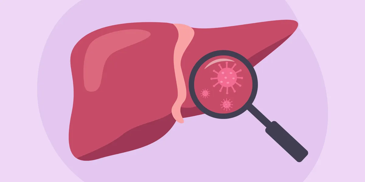
Facts About Liver Disease – HealthyWomen
May is Hepatitis Awareness Month. Around 4.5 million adults in the United States are living with chronic liver disease — and women are more likely than men to develop chronic liver disease. Some people may associate liver disease with alcohol use, but the truth is, many different factors, like excess weight, autoimmune conditions and even viruses, can cause liver disease. Certain types of liver disease also primarily target women. Here’s a closer look at your liver, what it does in your body and what can go wrong with it. Your liver: What it is and what it does All hail the liver, which, at around three pounds, is the largest organ located inside the body. (The skin is technically your largest organ.) The liver may not get as much attention as, say, your heart or kidneys, but the football-shaped, reddish-brown organ located just under your ribs performs hundreds of impressive feats daily. Plus, it’s the only organ inside your body that can heal itself and grow back if part of it is damaged or removed. Honestly, how cool is that? Here’s a brief snapshot of just some of the functions your liver performs on the regular: Processes nutrients from foods Produces vital proteins Filters toxins from blood (this role is not just for the kidneys!) Breaks down harmful substances Stores vitamins and minerals Clears out old red blood cells Makes the necessary components for your blood to clot The National Institutes of Health (NIH) describes the liver as “tough” and “resilient.” However strong the liver may be, it’s not indestructible. Even an organ capable of healing itself can get damaged, especially if it’s overworked long-term. What is liver disease? Liver disease can be acute (short-term) or chronic (long-term). Acute liver disease is rarer than chronic liver disease. In both cases, the liver is too overwhelmed to function, but they usually have different causes. Acute liver disease Short-term liver disease is typically caused by a viral infection, such as hepatitis A, or in some cases, an overdose of acetaminophen, also known as Tylenol. (Those label warnings on the bottle are there for a reason.) Depending on the extent of the damage, acute liver disease is often treatable, especially if caught early enough. In rare cases, acute liver disease can cause complete liver failure, and in that case, a liver transplant may be needed. Chronic liver disease Next up: Chronic liver disease, which is the more common condition. One of the key roles the liver plays is filtering toxins out of the blood, which makes it especially vulnerable to disease over time. If the liver is constantly exposed to high levels of toxins and forced to work at higher-than-normal loads, even the toughest of organs can wear down over time. There are four stages of chronic liver disease, which progress by severity: Hepatitis: This is the inflammation stage, when the liver is responding to injury or trauma. If no intervention occurs to stop the inflammation, it will progress to the next stage of fibrosis. Fibrosis: In this stage, as the liver is damaged, healthy tissue is replaced by scar tissue. The liver cannot function normally with bands of scarred tissue. However, even at this stage, if caught early enough, the scar tissue can be reversed. If the liver is not given the chance to heal, however, the next stage is permanent scarring. Cirrhosis: Once cirrhosis of the liver occurs, the scarring in the liver is usually permanent, although sometimes it can be reversible in its early stages if the underlying cause is treated. As liver function gets worse, symptoms in the body will gradually appear. However, your body is quite good at compensating when your liver isn’t working the way it should, so it can be years before noticeable symptoms appear. Even with permanent scarring, if the liver disease is caught fairly early on, it’s possible to slow down or stop further damage. Liver failure: The fourth and final stage of liver disease is liver failure. The disease still gets worse slowly, and symptoms can take some time to show, but eventually, full signs of liver failure will be apparent. The only treatment for liver failure is a liver transplant. Symptoms of liver disease If you have something acutely wrong with your liver, you’re more likely to experience extreme upper right abdominal pain, nausea and vomiting, and general feelings of being unwell. With chronic liver disease, you can have similar symptoms, but you can also experience worsening symptoms of: Jaundice (when the whites of your eyes and skin appear yellow) Confusion Itchy skin without a rash White poop Very dark urine Easy bruising and bleeding Small, yellow fat bumps on your skin or eyelids Weight loss Muscle loss Musty-smelling breath Difficulty digesting fats Swelling in your hands and feet Loss of menstrual cycle Swelling of the testes Chronic liver disease tends to get worse slowly over time, with the body making up for a lot of damage until the disease is very advanced. So people may not notice symptoms of liver disease right away. What causes liver disease? Liver disease can occur from excessive alcohol usage — and that’s probably the most commonly known reason. However, it can also occur without any direct lifestyle link. For instance, an infection or hereditary condition can lead to liver disease. There are over 100 different types of liver disease, but some of the most commonly seen include: Autoimmune liver disease: Autoimmune hepatitis, like other types of autoimmune diseases, can happen randomly when the body mistakes healthy liver cells as foreign cells and attacks them. This type of liver disease is more common in women and people assigned female at birth (AFAB). Autoimmune hepatitis can occur in middle-aged women (Type 1) or girls ages 2–14 (Type 2). Primary biliary cholangitis (PBC): PBC is another type of chronic liver disease that occurs mostly in women. It’s also thought to be related to the immune system and is most common in middle-aged women. Viral hepatitis: Viruses can
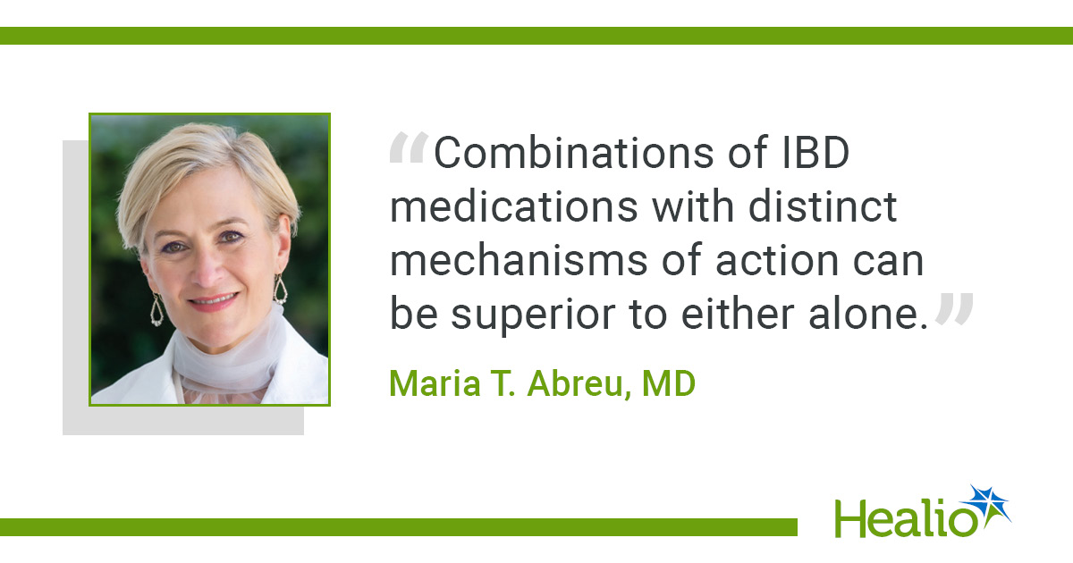
Co-antibody combo bests golimumab, guselkumab in refractory IBD
Add topic to email alerts Receive an email when new articles are posted on Please provide your email address to receive an email when new articles are posted on . “ data-action=”subscribe”> Subscribe We were unable to process your request. Please try again later. If you continue to have this issue please contact customerservice@slackinc.com. Back to Healio Key takeaways: Co-antibody combination therapy showed positive results in patients with IBD with inadequate response to two or more systemic therapies. At week 48, high dose co-antibody combination outperformed golimumab. CHICAGO — Combining golimumab and guselkumab into JNJ-78934804, a fixed-dose co-antibody therapy, helped overcome treatment failure in inflammatory bowel disease and exceeded dose-dependent benefits of either drug alone, according to data. Two parallel phase 2b trials combining the anti-tumor necrosis factor and anti-interleukin-23 medications in Crohn’s disease and ulcerative colitis, DUET-CD and DUET-UC, were presented by lead authors at Digestive Disease Week. “Results show that people with Crohn’s disease who have had inadequate response to two or more systemic therapies do strikingly better with combination therapy of two drugs with different mechanisms of action, specifically with golimumab, an anti-TNF antibody, and guselkumab, an anti-IL-23 antibody, rather than with treatment with either one alone,” Bruce E. Sands, MD, MS, chief of the Dr. Henry D. Janowitz Division of Gastroenterology at Mount Sinai Health System and lead author of the CD study, told Healio. Similar results were observed in the parallel UC trial, according to lead author Maria T. Abreu, MD, executive director at F. Widjaja IBD Institute at Cedars-Sinai Medical Center. “The study highlights that there is a path forward for UC patients who have been on more than two types of mechanisms of action of medications,” she told Healio. “Combining anti-TNF and anti-IL-23 was safe and showed superiority in the toughest to treat UC patients.” Crohn’s study results In the DUET-CD study, 693 adults with Crohn’s disease (57% men) were randomly assigned to receive guselkumab (Tremfya, Janssen), golimumab (Simponi, Janssen), low-, mid- or high-dose JNJ-78934804 (JNJ-4804, Johnson & Johnson) or placebo. Half of the patients had a prior inadequate response to two or more classes of systemic therapy. The coprimary endpoints were clinical and endoscopic remission at week 48, while other key endpoints included endoscopic remission, deep remission and corticosteroid-free clinical remission at that time point. High-dose JNJ-4804 achieved higher rates of the co-primary endpoints than golimumab, including clinical remission (difference = 25.7; P < .001) and endoscopic response (difference = 18.5; nominal P < .001), with similar or greater efficacy compared with guselkumab. The researchers also found that differences in treatment were greater among those who were refractory to two or more systemic therapy classes. At week 48, high-dose JNJ-4804 demonstrated clinically meaningful improvements in both clinical remission and endoscopic response compared with golimumab (27.3 and 21.2, respectively), guselkumab (21.2 and 11.7) and placebo (39.4 and 35). Exposure-adjusted safety events with JNJ-4804 were similar to or lower than those reported with placebo or golimumab. Most serious adverse events were gastrointestinal-related and serious infections were uncommon. Safety was comparable to that observed with monotherapies. This “fixed-dose combination of golimumab and guselkumab outperformed either one alone” in refractory Crohn’s disease without increased risk for adverse events compared with monotherapy, Sands told Healio. UC study results For the UC study, 572 individuals (57% men) were also randomly assigned to the same medications in the same way as the CD trial. Patients had a mean UC duration of 9 years (standard deviation, 7.5). Most patients had severe endoscopic disease, with 72% scoring 3 on the Mayo Endoscopy Subscore, 70% having modified Mayo scores of 7–9 and 45% reporting an inadequate response to two or more systemic therapy classes. Abreu and colleagues reported findings consistent with the DUET-CD study: High-dose JNJ-4804 was significantly superior to golimumab (28.4; P < .001) and comparable to guselkumab (6.3) in the study’s primary endpoint. At week 48, treatment responses were greater in patients who were refractory to two or more systemic therapy classes. In this subgroup, high-dose JNJ-4804 resulted in clinically meaningful improvements in clinical remission, corticosteroid-free clinical remission, endoscopic improvement and histologic remission and endoscopic improvement compared with golimumab (22.9, 20.5, 31.9, and 27.3, respectively), guselkumab (12.5, 12.3, 17.3, 16.9) and placebo (19.7, 17.2, 28.3, 23.6). Safety events per 100 patient-years in the JNJ-4804 groups were either comparable or lower than those observed in the other treatment groups. As in the DUET-CD study, most serious adverse events were GI-related with low rates of serious infection. “Combinations of IBD medications with distinct mechanisms of action can be superior to either alone,” Abreu told Healio. “The impact appears to be greatest for those patients who have already been on more than two types of therapy.” Both parallel trials are set to continue to phase 3 trials based on data, according to a press release on Sands’ and Abreu’s presentations. “These results suggest the power of combination therapy after attempts at treatment with single agents fail,” said Sands . For more information: Maria T. Abreu, MD, can be reached at maria.abreu@cshs.org. Bruce E. Sands, MD, MS, is Dr. Burrill B. Crohn Professor of Medicine at Icahn School of Medicine at Mount Sinai. He can be reached at bruce.sands@mssm.edu. Published by: Sources/Disclosures Source: Sands BE, et al. Efficacy and safety of the first co-antibody therapy, JNJ-78934804, in patients with moderately to severely active Crohn’s disease refractory to systemic therapies. Presented at: Digestive Disease Week; May 2-5, 2026; Chicago. Reference: Abreu MT, et al. Efficacy and safety of the first co-antibody therapy, JNJ-78934804, in patients with moderately to severely active ulcerative colitis refractory to systemic therapies. Presented at: Digestive Disease Week; May 2-5, 2026; Chicago. Disclosures: Healio was unable to confirm relevant financial disclosures at time of publication. Ask a clinical question and tap into Healio AI’s knowledge base. PubMed, enrolling/recruiting trials, guidelines Clinical Guidance, Healio CME, FDA news Healio’s exclusive daily news coverage of clinical