
Spinal cord stimulators, also known as neuromodulation devices, are medical devices that are designed to help manage chronic pain by interrupting pain signals before they reach the brain. These devices are typically implanted in the body and use electrical stimulation to alter the way that pain signals are sent through the spinal cord. What Are They Used For? Spinal cord stimulators are typically used to treat chronic pain conditions that have not responded to other treatments such as medication or physical therapy. They are often used to treat conditions such as chronic back pain, nerve damage, and other types of neuropathic pain. While spinal cord stimulators can be an effective treatment option for some patients, they are not right for everyone. Patients must undergo a thorough trial evaluation to determine if they are a good candidate for spinal cord stimulation. How Do They Work? Spinal cord stimulators work by interrupting the pain signals that are sent through the spinal cord. The device is designed to deliver electrical pulses to the nerves in the spinal cord, which can help to reduce the sensation of pain. These electrical pulses can also help to stimulate the release of endorphins, which are the body’s natural painkillers. The spinal cord stimulator device consists of several components, including a small generator that is implanted under the skin, a wire that is placed in the epidural space of the spinal cord, and a remote control that the patient can use to adjust the level of stimulation. The generator produces a mild electrical current, which is carried by the wire to the nerves in the spinal cord. The patient can adjust the level of stimulation using the remote control, which allows them to customize the level of pain relief they receive. What Are the Different Types of Spinal Cord Stimulators Available? There are several different types of spinal cord stimulators available, each with its own unique features and benefits. Some of the most common types of spinal cord stimulators include the following types: Conventional Spinal Cord Stimulators These are the most common type of spinal cord stimulators and are designed to deliver electrical pulses to the nerves in the spinal cord. High-Frequency Spinal Cord Stimulators These devices use a higher frequency of electrical stimulation than conventional spinal cord stimulators and are often used to treat more severe pain. Burst Spinal Cord Stimulators These devices deliver electrical pulses in bursts, which can help to reduce the sensation of pain without causing discomfort. Dorsal Root Ganglion (DRG) Stimulators These devices are designed to target specific nerves in the spinal cord and can be used to treat pain in specific areas of the body, such as the feet or hands. Peripheral Nerve Stimulators These devices are used to stimulate nerves outside of the spinal cord, such as those in the arms or legs. They can be used to treat chronic pain conditions in these areas. Pre-Surgery Trial Process Patient Evaluation: The patient undergoes a comprehensive evaluation by a pain management specialist to determine if they are a suitable candidate for spinal cord stimulator surgery. Pre-Trial Assessment: The patient undergoes imaging tests such as an MRI or CT scan to assess the spinal cord, nerves, and surrounding tissues. The specialist discusses the spinal cord stimulator surgery, including potential risks and benefits. Trial Implantation: A temporary electrode is placed near the spinal cord under local anesthesia, connected to an external device that sends electrical impulses. The patient keeps a pain diary for several days to a week. Post-Trial Evaluation: The patient meets with the specialist to evaluate the effectiveness of the spinal cord stimulator therapy in managing their pain. If significant pain relief is experienced, permanent implantation of the spinal cord stimulator device is recommended. What to Expect during a Spinal Cord Stimulator Implant Surgery The implantation surgery for a spinal cord stimulator typically takes a few hours to complete and is performed under general anesthesia. The surgeon will make a small incision in the patient’s back and place the generator device under the skin, usually in the lower abdomen or buttocks area. The wire lead will then be carefully guided into the epidural space of the spinal cord, with the guidance of X-ray imaging. Once the wire is in place, the surgeon will secure it in position and close the incision with stitches. After discharge, patients will need to take care to avoid bending or twisting their back for several weeks to allow the incision to heal. Patients may also be given a temporary external controller to use until the incision has fully healed and they are ready to use the permanent remote control. During the recovery period, patients may experience some discomfort or soreness at the incision site, which can be managed with pain medication. Patients will need to avoid activities that put excessive strain on their back, such as heavy lifting or strenuous exercise, for several weeks after the surgery. Are There Any Risks Involved? As with any surgical procedure, there are risks associated with the implantation surgery for a spinal cord stimulator. Some of the potential risks and complications include: ● Infection at the incision site or around the implanted device ● Bleeding or hematoma formation ● Pain or discomfort at the implantation site ● Device malfunction or failure ● Allergic reaction to the implant materials or anesthesia ● Nerve damage or spinal cord injury ● Displacement or migration of the implanted wire or device ● Spinal fluid leak or headache ● Movement restriction and loss of flexibility in the implanted area ● Need for device removal or revision surgery. It’s important to discuss the potential risks and benefits of spinal cord stimulator implantation surgery with your healthcare provider to determine if it is the right treatment option for you. They can also help you develop a plan to manage any potential risks and complications associated with the procedure. Whether you’re
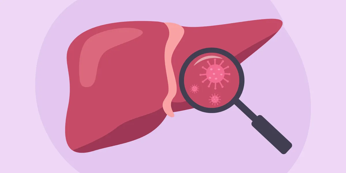
Facts About Liver Disease – HealthyWomen
May is Hepatitis Awareness Month. Around 4.5 million adults in the United States are living with chronic liver disease — and women are more likely than men to develop chronic liver disease. Some people may associate liver disease with alcohol use, but the truth is, many different factors, like excess weight, autoimmune conditions and even viruses, can cause liver disease. Certain types of liver disease also primarily target women. Here’s a closer look at your liver, what it does in your body and what can go wrong with it. Your liver: What it is and what it does All hail the liver, which, at around three pounds, is the largest organ located inside the body. (The skin is technically your largest organ.) The liver may not get as much attention as, say, your heart or kidneys, but the football-shaped, reddish-brown organ located just under your ribs performs hundreds of impressive feats daily. Plus, it’s the only organ inside your body that can heal itself and grow back if part of it is damaged or removed. Honestly, how cool is that? Here’s a brief snapshot of just some of the functions your liver performs on the regular: Processes nutrients from foods Produces vital proteins Filters toxins from blood (this role is not just for the kidneys!) Breaks down harmful substances Stores vitamins and minerals Clears out old red blood cells Makes the necessary components for your blood to clot The National Institutes of Health (NIH) describes the liver as “tough” and “resilient.” However strong the liver may be, it’s not indestructible. Even an organ capable of healing itself can get damaged, especially if it’s overworked long-term. What is liver disease? Liver disease can be acute (short-term) or chronic (long-term). Acute liver disease is rarer than chronic liver disease. In both cases, the liver is too overwhelmed to function, but they usually have different causes. Acute liver disease Short-term liver disease is typically caused by a viral infection, such as hepatitis A, or in some cases, an overdose of acetaminophen, also known as Tylenol. (Those label warnings on the bottle are there for a reason.) Depending on the extent of the damage, acute liver disease is often treatable, especially if caught early enough. In rare cases, acute liver disease can cause complete liver failure, and in that case, a liver transplant may be needed. Chronic liver disease Next up: Chronic liver disease, which is the more common condition. One of the key roles the liver plays is filtering toxins out of the blood, which makes it especially vulnerable to disease over time. If the liver is constantly exposed to high levels of toxins and forced to work at higher-than-normal loads, even the toughest of organs can wear down over time. There are four stages of chronic liver disease, which progress by severity: Hepatitis: This is the inflammation stage, when the liver is responding to injury or trauma. If no intervention occurs to stop the inflammation, it will progress to the next stage of fibrosis. Fibrosis: In this stage, as the liver is damaged, healthy tissue is replaced by scar tissue. The liver cannot function normally with bands of scarred tissue. However, even at this stage, if caught early enough, the scar tissue can be reversed. If the liver is not given the chance to heal, however, the next stage is permanent scarring. Cirrhosis: Once cirrhosis of the liver occurs, the scarring in the liver is usually permanent, although sometimes it can be reversible in its early stages if the underlying cause is treated. As liver function gets worse, symptoms in the body will gradually appear. However, your body is quite good at compensating when your liver isn’t working the way it should, so it can be years before noticeable symptoms appear. Even with permanent scarring, if the liver disease is caught fairly early on, it’s possible to slow down or stop further damage. Liver failure: The fourth and final stage of liver disease is liver failure. The disease still gets worse slowly, and symptoms can take some time to show, but eventually, full signs of liver failure will be apparent. The only treatment for liver failure is a liver transplant. Symptoms of liver disease If you have something acutely wrong with your liver, you’re more likely to experience extreme upper right abdominal pain, nausea and vomiting, and general feelings of being unwell. With chronic liver disease, you can have similar symptoms, but you can also experience worsening symptoms of: Jaundice (when the whites of your eyes and skin appear yellow) Confusion Itchy skin without a rash White poop Very dark urine Easy bruising and bleeding Small, yellow fat bumps on your skin or eyelids Weight loss Muscle loss Musty-smelling breath Difficulty digesting fats Swelling in your hands and feet Loss of menstrual cycle Swelling of the testes Chronic liver disease tends to get worse slowly over time, with the body making up for a lot of damage until the disease is very advanced. So people may not notice symptoms of liver disease right away. What causes liver disease? Liver disease can occur from excessive alcohol usage — and that’s probably the most commonly known reason. However, it can also occur without any direct lifestyle link. For instance, an infection or hereditary condition can lead to liver disease. There are over 100 different types of liver disease, but some of the most commonly seen include: Autoimmune liver disease: Autoimmune hepatitis, like other types of autoimmune diseases, can happen randomly when the body mistakes healthy liver cells as foreign cells and attacks them. This type of liver disease is more common in women and people assigned female at birth (AFAB). Autoimmune hepatitis can occur in middle-aged women (Type 1) or girls ages 2–14 (Type 2). Primary biliary cholangitis (PBC): PBC is another type of chronic liver disease that occurs mostly in women. It’s also thought to be related to the immune system and is most common in middle-aged women. Viral hepatitis: Viruses can
Co-antibody combo bests golimumab, guselkumab in refractory IBD
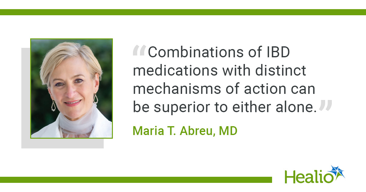
Add topic to email alerts Receive an email when new articles are posted on Please provide your email address to receive an email when new articles are posted on . “ data-action=”subscribe”> Subscribe We were unable to process your request. Please try again later. If you continue to have this issue please contact customerservice@slackinc.com. Back to Healio Key takeaways: Co-antibody combination therapy showed positive results in patients with IBD with inadequate response to two or more systemic therapies. At week 48, high dose co-antibody combination outperformed golimumab. CHICAGO — Combining golimumab and guselkumab into JNJ-78934804, a fixed-dose co-antibody therapy, helped overcome treatment failure in inflammatory bowel disease and exceeded dose-dependent benefits of either drug alone, according to data. Two parallel phase 2b trials combining the anti-tumor necrosis factor and anti-interleukin-23 medications in Crohn’s disease and ulcerative colitis, DUET-CD and DUET-UC, were presented by lead authors at Digestive Disease Week. “Results show that people with Crohn’s disease who have had inadequate response to two or more systemic therapies do strikingly better with combination therapy of two drugs with different mechanisms of action, specifically with golimumab, an anti-TNF antibody, and guselkumab, an anti-IL-23 antibody, rather than with treatment with either one alone,” Bruce E. Sands, MD, MS, chief of the Dr. Henry D. Janowitz Division of Gastroenterology at Mount Sinai Health System and lead author of the CD study, told Healio. Similar results were observed in the parallel UC trial, according to lead author Maria T. Abreu, MD, executive director at F. Widjaja IBD Institute at Cedars-Sinai Medical Center. “The study highlights that there is a path forward for UC patients who have been on more than two types of mechanisms of action of medications,” she told Healio. “Combining anti-TNF and anti-IL-23 was safe and showed superiority in the toughest to treat UC patients.” Crohn’s study results In the DUET-CD study, 693 adults with Crohn’s disease (57% men) were randomly assigned to receive guselkumab (Tremfya, Janssen), golimumab (Simponi, Janssen), low-, mid- or high-dose JNJ-78934804 (JNJ-4804, Johnson & Johnson) or placebo. Half of the patients had a prior inadequate response to two or more classes of systemic therapy. The coprimary endpoints were clinical and endoscopic remission at week 48, while other key endpoints included endoscopic remission, deep remission and corticosteroid-free clinical remission at that time point. High-dose JNJ-4804 achieved higher rates of the co-primary endpoints than golimumab, including clinical remission (difference = 25.7; P < .001) and endoscopic response (difference = 18.5; nominal P < .001), with similar or greater efficacy compared with guselkumab. The researchers also found that differences in treatment were greater among those who were refractory to two or more systemic therapy classes. At week 48, high-dose JNJ-4804 demonstrated clinically meaningful improvements in both clinical remission and endoscopic response compared with golimumab (27.3 and 21.2, respectively), guselkumab (21.2 and 11.7) and placebo (39.4 and 35). Exposure-adjusted safety events with JNJ-4804 were similar to or lower than those reported with placebo or golimumab. Most serious adverse events were gastrointestinal-related and serious infections were uncommon. Safety was comparable to that observed with monotherapies. This “fixed-dose combination of golimumab and guselkumab outperformed either one alone” in refractory Crohn’s disease without increased risk for adverse events compared with monotherapy, Sands told Healio. UC study results For the UC study, 572 individuals (57% men) were also randomly assigned to the same medications in the same way as the CD trial. Patients had a mean UC duration of 9 years (standard deviation, 7.5). Most patients had severe endoscopic disease, with 72% scoring 3 on the Mayo Endoscopy Subscore, 70% having modified Mayo scores of 7–9 and 45% reporting an inadequate response to two or more systemic therapy classes. Abreu and colleagues reported findings consistent with the DUET-CD study: High-dose JNJ-4804 was significantly superior to golimumab (28.4; P < .001) and comparable to guselkumab (6.3) in the study’s primary endpoint. At week 48, treatment responses were greater in patients who were refractory to two or more systemic therapy classes. In this subgroup, high-dose JNJ-4804 resulted in clinically meaningful improvements in clinical remission, corticosteroid-free clinical remission, endoscopic improvement and histologic remission and endoscopic improvement compared with golimumab (22.9, 20.5, 31.9, and 27.3, respectively), guselkumab (12.5, 12.3, 17.3, 16.9) and placebo (19.7, 17.2, 28.3, 23.6). Safety events per 100 patient-years in the JNJ-4804 groups were either comparable or lower than those observed in the other treatment groups. As in the DUET-CD study, most serious adverse events were GI-related with low rates of serious infection. “Combinations of IBD medications with distinct mechanisms of action can be superior to either alone,” Abreu told Healio. “The impact appears to be greatest for those patients who have already been on more than two types of therapy.” Both parallel trials are set to continue to phase 3 trials based on data, according to a press release on Sands’ and Abreu’s presentations. “These results suggest the power of combination therapy after attempts at treatment with single agents fail,” said Sands . For more information: Maria T. Abreu, MD, can be reached at maria.abreu@cshs.org. Bruce E. Sands, MD, MS, is Dr. Burrill B. Crohn Professor of Medicine at Icahn School of Medicine at Mount Sinai. He can be reached at bruce.sands@mssm.edu. Published by: Sources/Disclosures Source: Sands BE, et al. Efficacy and safety of the first co-antibody therapy, JNJ-78934804, in patients with moderately to severely active Crohn’s disease refractory to systemic therapies. Presented at: Digestive Disease Week; May 2-5, 2026; Chicago. Reference: Abreu MT, et al. Efficacy and safety of the first co-antibody therapy, JNJ-78934804, in patients with moderately to severely active ulcerative colitis refractory to systemic therapies. Presented at: Digestive Disease Week; May 2-5, 2026; Chicago. Disclosures: Healio was unable to confirm relevant financial disclosures at time of publication. Ask a clinical question and tap into Healio AI’s knowledge base. PubMed, enrolling/recruiting trials, guidelines Clinical Guidance, Healio CME, FDA news Healio’s exclusive daily news coverage of clinical

What Is Radiofrequency Nerve Ablation?
Are you experiencing chronic pain? Were such pain management methods as medications, physical therapy, or injections unsuccessful? Radiofrequency nerve ablation is a minimally invasive procedure that can help. Keep reading to find out more about this procedure. Overview of Radiofrequency Nerve Ablation Nerve fibers are responsible for carrying pain signals from inflamed or injured joints to the brain. Nerve ablation uses radio waves to destroy specific nerves in order to alleviate chronic pain. Most commonly, nerve ablation is used to treat pain associated with the joints of the spine, also known as facet joints. Facet joints are small joints located between the vertebrae of the spine and are responsible for allowing movement and stability in the spine. When Is Nerve Ablation Performed? The procedure is typically performed on patients who have not experienced relief from other pain management methods such as medications, physical therapy, or injections. The following conditions are typically treated using nerve ablation: Failed Back Surgery Syndrome—continued pain despite surgery to the spine Spondylosis—osteoarthritis causing degeneration of the facet joints Lumbago—lower back pain Cervicalgia—neck pain Scoliosis—abnormal curvature of the spine Radiofrequency ablation should not be performed on people who have an infection, are pregnant, or have bleeding problems. What Does the Procedure Involve? The nerve ablation procedure involves the use of special needles that are inserted through the skin under continuous X-ray guidance onto the site of the medial branch nerve. Once the needles are in place, a small electrical current is passed through them for a sensory test that causes discomfort, confirming that the needle is positioned correctly. Next, the needle tips are heated with a specialized machine to 90 degrees Celsius for 60 total seconds to destroy the nerve responsible for transmitting pain signals from the joint to the brain. The Benefits of Nerve Ablation The benefits of nerve ablation are numerous. First and foremost, it can provide significant pain relief for patients suffering from chronic pain associated with the facet joints. Nerve ablation can improve their quality of life and allow them to engage in activities that they may have previously avoided due to pain, such as exercising. Additionally, nerve ablation is a minimally invasive procedure that does not require a lengthy hospital stay or recovery period, which can be appealing to patients who are looking for a quick and effective pain management solution. How Long Do the Results Last? It is important to note that nerve ablation is not a permanent solution to chronic pain. While the procedure can provide significant pain relief for several months to a year or more, the nerves that were destroyed will eventually regenerate, and the pain may return. How to Choose a Pain Management Practice When considering nerve ablation as a pain management option, it is important to choose a pain management practice that is experienced and qualified to perform the procedure. Here are some factors to consider: Board Certification Ensure that the pain management practice is board-certified and has a team of qualified and experienced pain management specialists. At Pain Treatment Centers of America, we have dedicated physicians, state-of-the-art surgery centers, medication monitoring, CLIA-certified labs, and the most combined pain management experience of any practice in the region. You can take a closer look at the certifications of each of our doctors on our website. Expertise Look for a pain management practice that has extensive experience performing nerve ablation procedures specifically for the type of pain you are experiencing. Our team at Pain Treatment Centers of America has 300 years of combined experience in pain management. Reputation Ask for referrals from your primary care physician, friends, or family members who have had successful nerve ablation procedures in the past. You can also check online reviews and ratings to get a sense of the pain management practice’s reputation. Pain Treatment Centers of America is a BBB Accredited business, and you can read patient testimonials here. Technology Ensure that the pain management practice uses the latest technology and equipment to perform the nerve ablation procedure. Advanced technology can improve the accuracy and effectiveness of the procedure and reduce the risk of complications. All of our qualified doctors and staff members are equipped with cutting-edge technology to provide the best pain management treatments available. Communication Choose a pain management practice that communicates effectively with patients, listens to their concerns, and explains the procedure and potential risks in detail. The pain management practice should be able to answer your questions and provide clear instructions on how to prepare for the procedure and what to expect during and after the procedure. We have detailed overviews of the following procedures: radiofrequency nerve ablation and knee joint nerve ablation so that you are fully prepared when you arrive at your appointment. Location and Availability Consider the location of the pain management practice and the availability of appointments that fit your schedule. Choosing a practice that is conveniently located and has flexible scheduling options can reduce the stress of the procedure and make it easier to follow up with your pain management specialist after the procedure. For your convenience, we at Pain Treatment Centers of America have fifteen locations across Arkansas, Mississippi, and Texas. In Conclusion Nerve ablation is a minimally invasive procedure that can provide significant pain relief for patients suffering from chronic pain associated with the facet joints of the spine. While the procedure is not a permanent solution, it can provide relief for several months to a year or more, allowing patients to return to their normal activities and avoid more invasive procedures such as surgery. As with any medical procedure, it is important to choose a qualified and experienced medical professional to perform the procedure and to work closely with your healthcare provider to determine the best pain management approach for your individual needs. If you would like to know whether the radiofrequency nerve ablation procedure is right for you, feel free to call us for more information at (844) 215-0731 today. At Pain Treatment Centers of America, we are

Minimally Invasive Surgery, Robotic-Assisted Procedures for Lung Cancer
Learning you might have a disease like lung cancer is scary. Luckily, there are surgical options for diagnosis and treatment that are minimally invasive. This means they only involve tiny cuts, and you recover much more quickly compared to traditional, or open, surgery. Understanding a few different types of minimally invasive procedures, and how they help people with lung cancer and other conditions, may help you feel less anxious about what you’re facing. Common minimally invasive procedures One minimally invasive procedure that is used to diagnose and treat a wide variety of health problems is an endoscopy. During an endoscopy, your healthcare provider (HCP) puts an endoscope, which is a long, thin tube, inside your body to get a close-up view of the body part they need to check. This tube usually has a light and a camera at the end of it, and your HCP looks at a screen that shows them what the camera is seeing. There are many different types of endoscopies, but they all work in the same basic way. A type of endoscope called a laparoscope can be used with surgical tools in a minimally invasive surgery called a laparoscopy to check for problems in your stomach or pelvic area. During a laparoscopy, the laparoscope goes in your stomach through a small cut and displays images on a monitor for your surgeon to view. Robotic-assisted surgery is another minimally invasive procedure. It involves a high definition (HD) camera that shows a close up of the area and a robotic arm with tiny surgical tools at the end. There’s also a control panel that looks like a video game joystick. The surgeon controls the robotic arm, which is a type of surgical instrument, to perform the surgery. When are minimally invasive procedures used? While there will be times when more traditional procedures are needed, minimally invasive procedures can be used for many different health issues all over the body — including lung cancer and other health problems that affect the lungs. For example, HCPs may perform a type of endoscopy called a bronchoscopy that uses a tube to look inside your lungs and airways. This procedure can be used to check for lung cancer and to figure out how serious it is. Robotic bronchoscopy, which is done on systems like Ion and Monarch, involves a smaller tube and a control panel that an HCP uses to move the tube in precise ways and reach parts of the lung that a traditional bronchoscopy cannot reach. A 3D map of the lungs allows the HCP controlling the tube to see exactly where the tube is and where it needs to go, guiding the tube to difficult-to-reach nodules for biopsying. Minimally invasive procedures can also be used to treat lung cancer and other lung problems. Instead of the traditional approach, which is open surgery called a thoracotomy, minimally invasive procedures use smaller incisions and often offer shorter recovery times. With video-assisted thoracoscopic surgery (VATS), often used for small, early-stage lung cancer, your HCP makes a few small cuts in your chest, then uses a camera and special long-handled tools to perform the surgery while looking at a video screen. With robotic-assisted surgery, such as the da Vinci Surgical System or the Mako system, robotic arms (fully controlled by the doctor) are used in a minimally invasive way to treat more complex lung issues. Robotic-assisted surgery uses a video screen with high-definition 3D imaging. Perks of minimally invasive procedures Minimally invasive procedures only need small cuts so they’re much easier on the body, which may mean less pain for the patient, shorter recovery times and smaller scars. In addition, studies have shown that robotic surgery is associated with better outcomes than other types of minimally invasive surgery or open surgery both during and after surgery. The benefits of robotic surgery include reduced need for blood transfusions, lower rates of complications, shorter hospital stays, fewer hospital readmissions and even lower mortality rates. Surgery that takes less of a toll may be particularly helpful for people going through cancer treatment. When your body doesn’t have to work as hard to heal from surgery, it may be better able to handle what comes next in your treatment plan. And this kind of minimally invasive cancer treatment is on the rise. A recent study led by the Duke University School of Medicine that looked at over 76,000 lung cancer cases found lung cancer surgery is moving in a less invasive direction — particularly for younger and healthier patients. “This is a very encouraging finding for the entire community of professionals caring for lung cancer patients,” study author and Duke Medical School professor Xiaofei Wang, Ph.D., said in a press release. Know your options Whether you’re dealing with the scary possibility of a lung cancer diagnosis or another health problem entirely, a minimally invasive procedure may be a potential treatment tool. To learn more about your choices when it comes to less invasive treatments, talk to your HCP. They can walk you through your options and help you choose the best one for your unique needs. This educational resource was created with support from Intuitive. From Your Site Articles Related Articles Around the Web Source link

Haven’t applied AI in practice yet? ‘Play with it in everyday life first’
May 21, 2026 42 min watch Add topic to email alerts Receive an email when new articles are posted on Please provide your email address to receive an email when new articles are posted on . ” data-action=subscribe> Subscribe We were unable to process your request. Please try again later. If you continue to have this issue please contact customerservice@slackinc.com. Back to Healio Key takeaways: Using AI outside the clinic can help doctors get acquainted with it before applying it to clinical situations. AI models are useful for different types of tasks depending on how they were trained. Doctors who have not yet started using AI tools should first get comfortable using them in everyday life before adopting them in clinical practice. That is the advice from optometrist Scott A. Edmonds, OD, FAAO, a member of the Healio AI Advisory Board who has been using AI in various areas of his practice. “You learn to use the tool in a much less stressful environment,” Edmonds said. “You learn how the model works and how it thinks. … Then when I go to use it in the real world, in the clinic, I’m more attuned. I’m not afraid of it. I know what to ask it. “Play with it in everyday life first, and then take your knowledge base, go into the clinic and use it.” Edmonds joined Hansa Bhargava, MD, Healio’s chief clinical strategy and innovation officer, in a Healio Community webinar focused on AI in health care. He explained that his practice had not adopted AI until a patient who was a specialist in large language models, the technology behind services such as ChatGPT, explained how it could be beneficial. “She started telling me her background and asking me why we didn’t use [AI] in our practice. And I was like, ‘Well, I’m not really sure how,’” Edmonds said. “She gave me CliffsNotes, and within a week, we started to use it. Within a month, we were using it every single day, especially the AI scribes, which were just really a no-brainer for the kind of work we do in terms of capturing clinical data and putting it into a format that’s medically and legally easy.” Since then, his office’s use of AI has expanded. One way he frequently uses large language models is to keep up with the potential side effects and drug interactions of medications patients are taking, particularly ones newly approved by the FDA. “[Patients] expect you to know. And unless you’re using this kind of model, you can’t keep up,” he said. “That’s very helpful in everyday clinical practice because a day doesn’t go by there isn’t a new drug that somebody’s asking me about.” Edmonds explained that different AI chatbots are better for different purposes. He described ChatGPT as a “Swiss Army Knife of AI” trained on a wide variety of information, peer reviewed or not. On the other hand, products such as Healio AI ingest a more targeted range of fact-checked information and could be more appropriate for use in health care. Still, Edmonds cautioned that the answers from AI cannot necessarily be trusted. “It isn’t the end-all be-all. It has to be just a piece of information,” he said. “It’s only as good as, A, the database and, B, how you prompted it.” For more information: Hansa Bhargava, MD, can be reached at primarycare@healio.com. Scott A. Edmonds, OD, FAAO, can be reached at optometry@healio.com. Published by: Sources/Disclosures Source: Webinars Disclosures: Bhargava is Healio’s chief clinical strategy and innovation officer. Edmonds reports no relevant financial disclosures. Ask a clinical question and tap into Healio AI’s knowledge base. PubMed, enrolling/recruiting trials, guidelines Clinical Guidance, Healio CME, FDA news Healio’s exclusive daily news coverage of clinical data Learn more Add topic to email alerts Receive an email when new articles are posted on Please provide your email address to receive an email when new articles are posted on . ” data-action=subscribe> Subscribe We were unable to process your request. Please try again later. If you continue to have this issue please contact customerservice@slackinc.com. Back to Healio Source link

Growing Beyond the Lines I Thought Pain Drew: What Leading a Daily Support Group Has Taught Me
By Michele Rice Living with chronic pain has a way of quietly drawing lines around your life. Some of those lines are physical. Some are emotional. Some are routines we build to survive the day. Over time, those lines can start to feel permanent—like invisible boundaries that define what we can and cannot do. For me, mornings have always been one of those lines. I’m not someone who jumps out of bed ready to start the day. It takes me a long time to get moving. My body is stiff, my pain is loud, and my brain feels slow to wake up. I also tend to resist change. I like knowing what to expect and staying within the routines that help me manage my health. So if you had asked me not long ago whether I would ever lead a chronic pain support group that meets in the morning every day, I would have said, “No way.” And yet… Here I am. A Decision Made From the Heart I recently learned that the U.S. Pain Foundation’s daily peer support group needed a new leader. For many people living with chronic pain, these groups are more than just a meeting on a calendar. They are a lifeline, a place where people understand each other in ways the outside world often cannot. In that moment, I didn’t stop to analyze whether I was capable of doing it. I didn’t weigh the pros and cons. I didn’t even really think about how hard it might be. All I thought about was the people in that group. I didn’t want them to face each morning without the place they relied on for connection and support. So I stepped forward. At the time, I believed I was doing it for them. What I didn’t realize was how much it would change me, too. Pushing Past My Own Boundaries Leading the group every weekday morning has not been easy. It means getting up earlier than my body prefers. It means showing up on camera even when pain or fatigue are loud that day. It means being present and prepared to guide conversations when my instinct might be to stay quiet and conserve energy. In many ways, it pushed me outside the lines I thought my life had to stay within. And honestly? At first, it felt uncomfortable. But something unexpected happened as the weeks went on. Instead of feeling like something that was draining me, the group began filling me up. The Joy I Didn’t Expect There is something incredibly powerful about gathering with people who truly understand what it means to live with chronic pain. Every morning, people show up with honesty, humor, vulnerability, and resilience. Some days we talk about the difficult realities of living with pain. Other days we laugh. Often we learn from each other in ways that no textbook or medical appointment could ever teach us. I started the group thinking I was offering support. But what I discovered is that support flows in every direction. The people who attend the group have lifted me up just as much as I hoped to lift them. On days when my own pain feels heavy, their courage reminds me that none of us is carrying this alone. Finding Community in Unexpected Ways Another gift that came from stepping into this role has been the deepened connections with people who have helped along the way. Leading a group like this is not something you do alone. There are people who check in, help facilitate topics and conversations, offer encouragement, and simply show up day after day. Through this experience, I’ve built stronger bonds with people who share not just similar challenges, but similar compassion for others living with chronic pain. That kind of connection is rare and incredibly meaningful. Growing Outside the Lines Chronic pain often teaches us to live carefully. We learn our limits, adjust our routines, and try to protect the energy we have. Those boundaries are important. But sometimes we discover that the lines we thought were permanent are actually more flexible than we imagined. Stepping into this role showed me that growth can still happen, even in a life shaped by chronic illness. It reminded me that purpose can show up in unexpected ways. And it taught me that sometimes the things we do to help others end up helping us just as much. Or maybe even more. If there’s one thing this experience has taught me, it’s this: Chronic pain may change the shape of our lives, but it does not erase our ability to grow, connect, or contribute. Sometimes growth doesn’t look like climbing a mountain or running a marathon. Sometimes growth looks like logging on to a morning support group, turning on your camera, and saying, “Good morning, everyone.” And discovering that in doing so, you’ve stepped just a little bit beyond the lines you once thought you were stuck inside. —by Michele Rice Source link

Gout Diet 101 – HealthyWomen
May 22 is National Gout Awareness Day. Pop quiz: What is gout? Is it … The universal nickname for golden trout fish A trendy lip-plumping serum Weird Al Yankovic’s parody of the iconic 80s banger “Shout” A disease caused by high levels of uric acid in the blood The answer — unfortunately — is D. Gout is a type of inflammatory arthritis that causes pain and swelling in the joints. It develops when high levels of uric acid build up in your blood and form sharp needle-shaped crystals, called tophi, in and around joints. Yes, it’s as painful as it sounds. Although gout is more common in men than women, cases of gout have more than doubled among women in the past 20 years. Research is ongoing as to why this is, but we do know that certain lifestyle factors, including what you eat and drink can help manage levels of uric acid in the blood, lower the risk for flares and even stop flares from happening. Read: What You Need to Know About Gout in Women >> How diet affects gout Your body makes uric acid after breaking down a chemical called purine. Purine, which is produced in the body and also found in some foods and drinks, gets broken down into uric acid and then leaves the body in urine. You can get gout if your body is producing too many purines and, therefore making too much uric acid, or if your kidneys are not functioning well enough to remove the uric acid — or a combination of both. This is where your meals come in. Foods and beverages high in purines increase the amount of uric acid in the body, which makes it harder for the body to get rid of it and increases the risk of build-up. Foods to avoid when you have gout As a rule, purine is highest in animal-based products and beer. Foods and beverages to limit and/or avoid if you have gout include: Organ meats: Sorry, liver-and-onions lovers. Organ meats including tongue, kidneys and sweetbreads are very high in purines and should be avoided. Red meat: Beef, lamb and pork are high in purines, so it’s a good idea to treat meat like an occasional treat if you have gout. Some seafood: Certain types of seafood are higher in purines than others. Top offenders: mussels, scallops, trout, tuna, anchovies, sardines, shrimp and lobster. Alcohol: Drinking any type of alcohol — especially beer — increases uric acid production and slows kidney function, which can lead to a build-up and increase the risk for flares. Sugary food and drinks: Sugar and high-fructose corn syrup found in many different foods and beverages (soda, fruit juice, cereal, desserts, etc.) speed up the production of uric acid. What to eat when you have gout There are foods that can help lower uric acid, reduce inflammation and help prevent gout flares. Overall, this means eating more plant-based foods as a part of a healthy lifestyle. Low-purine foods can include: Non-meat proteins like tofu Low-fat and nondairy products Lean proteins like chicken (in moderation) Nuts, nut butter and whole grains Foods high in vitamin C can help decrease uric acid levels and help prevent gout attacks. Fruits like grapefruit, oranges, pineapples, kiwifruit, strawberries and peppers are all good options that are high in vitamin C but lower in fructose. And while we’re on the subject of fruit, tart cherries and tart cherry juice contain antioxidants that have anti-inflammatory properties and may lower the risk of gout attacks. Dietary choices can make a difference Your body naturally produces most of the uric acid in your blood, but what you eat and drink can play a role in managing gout flares. One recent study found eating plants emphasizing plant-based protein like legumes, nuts, seeds and whole grains lowers uric acid more than diets low in carbohydrates or unsaturated fat. If you’re living with gout, talk to your healthcare provider about finding an eating plan that can help prevent painful flares and keep uric acid levels in check. From Your Site Articles Related Articles Around the Web Source link

RFK Jr. fires vice chairs of USPSTF
Add topic to email alerts Receive an email when new articles are posted on Please provide your email address to receive an email when new articles are posted on . “ data-action=”subscribe”> Subscribe We were unable to process your request. Please try again later. If you continue to have this issue please contact customerservice@slackinc.com. Back to Healio Key takeaways: Two USPSTF leaders were fired after being told they had been “inappropriately appointed.” Experts expressed concerns over the future of the task force and how its instability could impact public health. HHS Secretary Robert F. Kennedy Jr. has fired both vice chairs of the U.S. Preventive Services Task Force, according to letters obtained by Healio. The vice chairs were Esa M. Davis, MD, MPH, FAAFP, a professor at the University of Maryland School of Medicine, and John B. Wong, MD, MACP, a professor at Tufts University School of Medicine. According to the task force’s website, Davis was appointed vice chair in March 2025. She previously served as a member from January 2021 to December 2024. Wong was appointed vice chair in March 2024. He previously served as a member from March 2018 to December 2023. Davis was supposed to serve as vice chair until March 13, 2027, and then chair until March 13, 2028, according to her appointment letter. Wong’s term was set to end in March 2027, according to reporting by Reuters. Alex H. Krist, MD, MPH, a former USPSTF member, told Healio that the task force now consists of eight members, although no chairs or vice chairs remain. The task force usually consists of 16 members, but it has been a while since new members were appointed. Firings followed a review of appointments In the letters, dated May 11, Kennedy said that he ordered a review of current USPSTF appointments “to ensure clarity, continuity and confidence in the [HHS’] exercise of its appointment and supervisory responsibilities, and to protect the integrity of the Task Force’s work.” He said that Davis’ and Wong’s firings, which are effective immediately, were a result of the review and done “to avoid uncertainty that could jeopardize the validity of future Task Force actions.” The terminations are not reflective of Davis’ and Wong’s years of service or performance on the task force, Kennedy added, but that HHS “is taking this step to help protect the Task Force and preserve confidence in the continuity and durability of its work.” Kennedy encouraged Davis and Wong to reapply for USPSTF membership during the nomination cycle ending on May 23 as their “continued participation would be highly valued.” Wong told Healio that the firing was a surprise, but that he and Davis submitted requests to rejoin. “We believe very much in the work of the task force. Our goal is to help and keep people healthy to live longer, healthier lives,” he said. When Wong asked if they could restore their chair statuses, he was told that they would only be considered for member roles, not leadership roles. ‘Inappropriately appointed’ In a letter to Roger D. Klein, MD, JD, director of the Agency for Healthcare Research and Quality, and Benjamin Robles, HHS deputy general counsel, Davis and Wong said they were told they had been “inappropriately appointed” during a meeting on May 12, which would make their chair tenures “invalid.” “Importantly, Secretary Kennedy’s termination letters neither state nor imply that our original appointments were somehow unlawful or inappropriate,” Davis and Wong wrote. “To the contrary, he implicitly confirms our validity by thanking us for our ‘many years of dedicated service to the Task Force’ which, by definition, would include January 2025 through the present.” When Davis and Wong asked for further clarification, “the response Dr. Klein provided to us was that our specific questions implicate legal and policy judgments that would not be appropriate to address in external correspondence,” Wong said. “The external part is that we’re no longer part of the task force, so it remains unknown to us exactly why in this context. But I can say the timing of this coincides with a solicitation for new task force members,” Wong said. HHS did not respond to Healio’s questions about why Davis and Wong were let go and whether the agency believes their appointments were in fact inappropriate. Kennedy not wanting Davis and Wong in leadership roles “is one of our areas of confusion,” Wong said. “There are some very nice compliments about early contributions and our expertise. He even says they’re deeply valued,” Wong said. Criticism from medical societies Uncertainty surrounded the USPSTF for months. Under Kennedy’s tenure, three task force meetings have been canceled since July 2025, and the USPSTF has published far fewer recommendations than under prior administrations. AMA President Bobby Mukkamala, MD, said his organization “is extremely concerned” about the terminations. “Today’s changes were foreshadowed by the earlier dismantling of the Advisory Committee on Immunization Practices,” he said in a statement. “We strongly urge HHS to restore the USPSTF’s long-standing, transparent process for selecting members, specifically clinicians with expertise in the fields of preventive medicine and primary care.” Mukkamala also urged HHS “to commit to once again holding regular Task Force meetings to ensure its important work can continue without further delay. Our patients’ lives depend on it.” ACP President Jan K. Carney, MD, MPH, MACP, said in a statement that Davis and Wong “serve as important representatives for internal medicine physicians and the patients that we care for. Both physicians are highly qualified experts, and we take issue with the lack of transparency in any review that Secretary Kennedy has conducted of members of the task force.” “The firings come as the task force has not met over the course of the past year and has been prevented from doing their work to ensure that the American public has up-to-date guidance, based on the best-available evidence, about preventive health care services. The USPSTF guidance is critical to a

What Is Sacroiliac Joint Fusion?
The sacroiliac joint is an important part of the body, connecting the spine to the pelvis. However, this joint can sometimes become stiff and painful, leading to a condition known as sacroiliac joint dysfunction. In extreme cases, a surgical procedure known as sacroiliac joint fusion may be recommended to relieve pain and restore mobility. Read on to learn more about what sacroiliac joint fusion is, who it can help, how it works, and who can provide it. What Is the Sacroiliac Joint? The sacroiliac joint (SI joint) is a small joint located between the sacrum (the triangular bone at the base of the spine) and the ilium (part of the pelvic bone). It is responsible for transmitting forces between the spine and the legs, providing stability, and absorbing shock. The SI joint is held together by strong ligaments and is reinforced by muscles in the surrounding area. Unfortunately, the SI joint can become painful due to a variety of reasons, including trauma, arthritis, or pregnancy. Pain in this area can be debilitating, making it difficult to perform everyday tasks such as walking or sitting for prolonged periods. In these cases, sacroiliac joint fusion surgery may be necessary to relieve the pain and restore function. Sacroiliac joint fusion involves the fusion of the sacrum and the ilium to create a solid connection between the two bones, thereby eliminating motion at the joint and reducing pain. The surgery can be performed using minimally invasive techniques, resulting in less trauma to the surrounding tissues and a quicker recovery time. How Does Sacroiliac Joint Fusion Surgery Work? Sacroiliac joint fusion surgery involves permanently joining the two bones that make up the sacroiliac joint. The surgery is performed under general anesthesia, and a small incision is made near the affected joint. During the procedure, bone grafts are placed between the sacrum and ilium bones to encourage the growth of new bone tissue. In some cases, metal screws or rods may be used to hold the bones in place until the fusion is complete. Sacroiliac joint fusion surgery has a high success rate and can provide relief for patients who have been suffering from chronic pain in the sacroiliac joint. However, it is important to talk to your doctor to determine if you are a candidate for the surgery and to discuss any potential risks or complications. Am I a Candidate for Sacroiliac Joint Fusion Surgery? If you have been experiencing chronic lower back pain that has not been relieved through non-surgical methods such as physical therapy, medications, or injections, then you may be a candidate for sacroiliac joint fusion surgery. This surgery is typically reserved for patients who have tried multiple other treatments and are still experiencing significant pain and discomfort. It is important to note that sacroiliac joint fusion is not a first-line treatment and is usually only recommended after other options have been exhausted. In order to determine if you are a good candidate for the procedure, your doctor will likely perform a physical exam and may order imaging tests such as X-rays or an MRI. They will also consider your medical history and any previous surgeries you may have had. It is important to have an open and honest conversation with your doctor about your symptoms and concerns, as well as the potential risks and benefits of the surgery. They can help you make an informed decision about whether sacroiliac joint fusion is right for you. How Long Does It Take To Recover from Sacroiliac Joint Fusion Surgery? Recovery from sacroiliac joint fusion surgery varies depending on the individual patient and the severity of the condition being treated. In the first few weeks after surgery, patients may experience some discomfort, swelling, and stiffness. Pain medication may be prescribed to help manage any discomfort. It’s important to follow your surgeon’s post-operative instructions closely to ensure proper healing. Physical therapy may also be recommended to help you regain strength, flexibility, and range of motion. Your surgeon may also recommend certain restrictions on activities, such as bending or lifting, to allow the joint to heal properly. Most patients can return to light activities within 2-4 weeks after surgery, but it can take several months to fully recover. During this time, it’s important to continue following your surgeon’s instructions, attend all follow-up appointments, and report any concerns or changes in your condition to help with healing. Are There Any Risks Associated with Sacroiliac Joint Fusion Surgery? Like any surgery, there are potential risks and complications associated with sacroiliac joint fusion. However, the risks associated with this procedure are relatively low compared to other surgeries. The most common risks include infection, bleeding, and nerve damage. Your surgeon will take steps to minimize these risks, such as using sterile techniques during the surgery and monitoring your nerve function closely. Another risk is that the fusion may not be successful, which means that the joint may not fuse properly or may not fuse at all. In some cases, additional surgery may be necessary to correct this issue. It’s also possible to experience pain or discomfort during the recovery period, although this can usually be managed with pain medication and other non-surgical treatments. Overall, sacroiliac joint fusion is a safe and effective option for patients who have not found relief from other treatments. Your surgeon will carefully evaluate your individual case and discuss the potential risks and benefits with you in detail before recommending this procedure. If you’re considering sacroiliac joint fusion surgery or have questions about the process, our compassionate experts at Pain Treatment Centers of America can provide guidance and support. Contact us today at (844) 215-0731 to learn more. Source link