
Diagnosing hair loss can be challenging. Many hair disorders share overlapping clinical features, and subtle differences in history, examination, trichoscopy, and sometimes biopsy are required to arrive at the correct diagnosis. As a result, several hair loss conditions are commonly misdiagnosed in everyday clinical practice. One of the most frequent diagnostic errors occurs when telogen effluvium (TE) or chronic telogen effluvium (CTE) is diagnosed in patients who actually have early androgenetic alopecia (AGA). Patients with early AGA often present with increased shedding, which can easily mimic telogen effluvium. However, careful examination frequently reveals early miniaturization of follicles, particularly along the central scalp or frontal region. Another condition that is frequently misunderstood is short anagen syndrome. Women who report that their hair never seems to grow long are sometimes incorrectly given this diagnosis. In reality, many of these patients have androgenetic alopecia, where progressive follicular miniaturization leads to shorter and finer hairs over time. True short anagen syndrome is relatively uncommon, and distinguishing it from other causes of reduced hair length requires careful evaluation. There is also frequent confusion between short anagen syndrome and loose anagen syndrome. These are distinct conditions with different mechanisms. Alopecia areata incognita is another diagnosis that is often applied too liberally. This condition presents with diffuse shedding and can resemble telogen effluvium. However, most patients referred to me with a presumed diagnosis of alopecia areata incognita ultimately do not have the condition. While certain trichoscopic findings may raise suspicion, a scalp biopsy is generally required to confirm the diagnosis. Finally, fibrosing alopecia in a pattern distribution (FAPD) is commonly misdiagnosed. Many clinicians incorrectly assume any LPP patient with androgenetic alopecia (AGA) should be laboratory as FAPD. FAPD is a special presentation. Careful history, clinical examination, trichoscopy, and occasionally biopsy are essential tools in avoiding these common diagnostic pitfalls. Source link

Low Level Laser Therapy (LLLT) for Hair Loss: Which Lasers are Better — Donovan Hair Clinic
I enjoyed discussions around the topic of low level laser devices for treating hair loss. Low-level light therapy (often called LLLT or “photobiomodulation”) is a non-drug option for androgenetic alopecia that uses red light (roughly 620–670 nm) to stimulate hair follicles. Two recent systematic reviews (one if which was also a meta-analysis) of randomized controlled trials (RCTs) concluded that the home-use devices can meaningfully increase hair density compared with sham (placebo-like) devices. But how do various types of devices- including laser diode vs LED compare? Nowadays, some devices are based on laser diodes, some LED and some have both!! Let’s review two important recent systematic reviews. First, some definitions! Laser vs LED — what do these terms mean? * Laser (LD = laser diode): emits a highly collimated, narrow-band beam (very “focused” light at a fairly specific wavelength). * LED (light-emitting diode): emits non-coherent, broader-band light that spreads more. Biologically, both aim to trigger follicle “energy” pathways: red light is absorbed by cytochrome-c oxidase in mitochondria, increasing ATP/ROS signaling that can promote cellular activity supportive of hair growth. What the studies show (and which is better)?? Across 7 double-blind RCTs (607 participants), laser therapies improved hair density versus sham with an overall standardized mean difference (SMD) ~1.27. When trials/devices were grouped by light source (LD vs LED) both reviews found a statistically significant difference favoring laser diodes alone over mixed LED+laser devices: * LDs alone: SMD 1.52 (95% CI 1.16–1.88) * LEDs + LDs: SMD 0.85 (95% CI 0.55–1.16) (p=0.043). Based on current RCT level evidence, laser-diode based devices seem a bit better. That said, these are not perfect head-to-head comparisons, follow-up is generally short (≤26 weeks), and LED-only data are limited References 1. Lueangarun S et al. J Clin Aesthet Dermatol. 2021;14(11):E64–75. 2. Gentile P, Garcovich S. Facial Plast Surg Aesthet Med. 2024;26(2) #lllt #hairloss #laser #androgeneticalopecia Source link
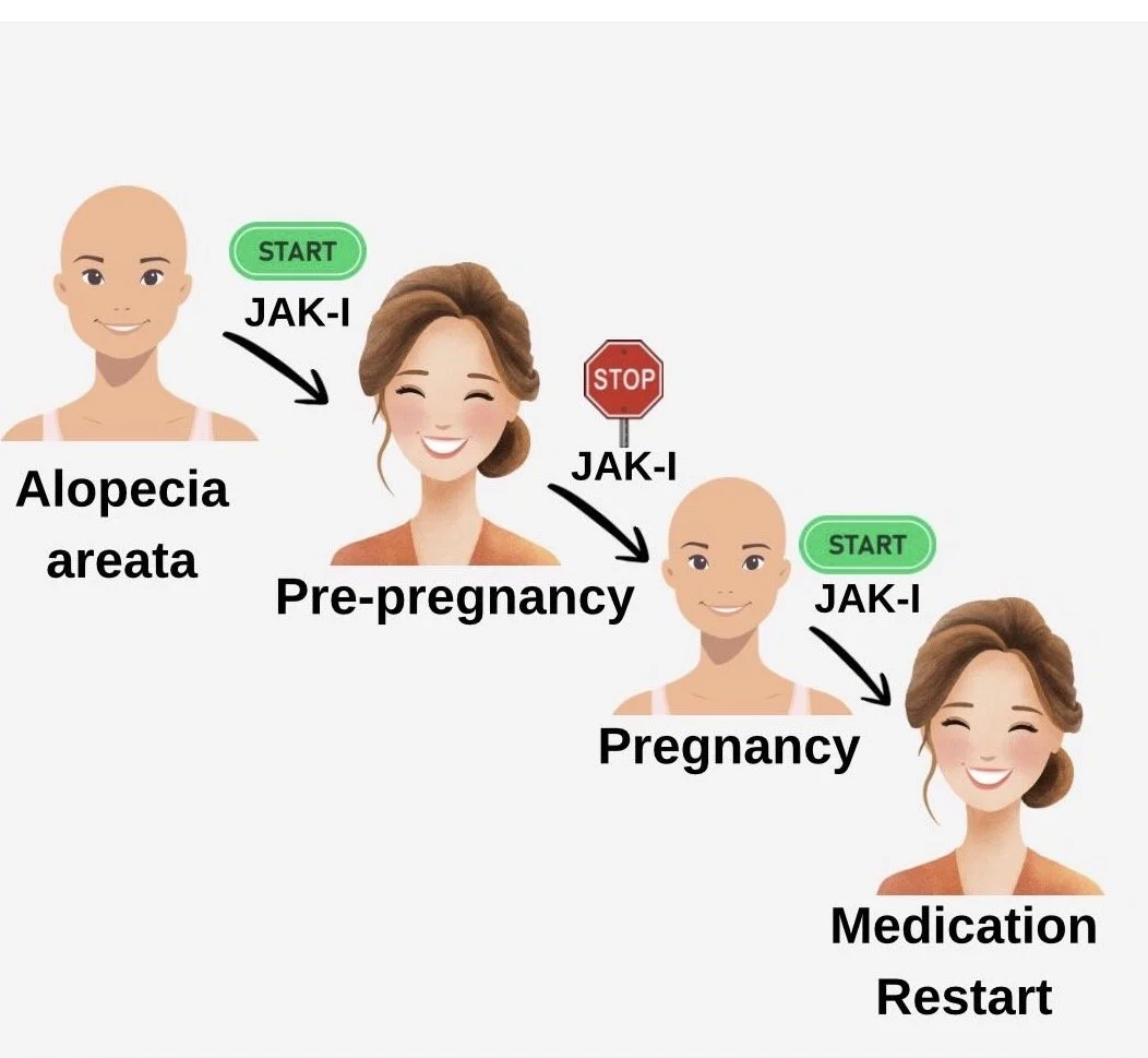
Stopping JAK Inhibitors in Pregnancy — Donovan Hair Clinic
Janus kinase inhibitors (JAK inhibitors) are medications that can help some patients with severe alopecia areata (AA) regrow hair. But because safety in pregnancy isn’t fully known, current recommendations are for all women stop treatment when trying to conceive. Unfortunately, this usually leads to hair loss. A nicely conducted new study set out to describe what happens to scalp hair when women stop JAK inhibitors for pregnancy — and what happens after restarting them postpartum. Researchers followed 9 women with severe AA through 14 pregnancies. All stopped oral JAK inhibitors before or early in pregnancy. RESULTS Every woman experienced significant hair loss after stopping treatment. However, once JAK inhibitors were restarted — usually within 4 months after delivery — patients regrew their hair, often back to or better than before. CONCLUSIONS This study is great news for the many women with alopecia areata who want to become pregnant but are terrified to stop their JAK inhibitor. Women with AA who must stop JAK inhibitors during pregnancy should be counseled that hair shedding is highly likely. The encouraging news is that restarting treatment after delivery (and after breastfeeding is done) led to meaningful regrowth in all cases. This study highlights the emotional burden these women face and the need for clearer pregnancy-related treatment guidelines in AA. REFERENCE Ogbutor C et al. Int J Womens Dermatol. 2025;11:e218 Source link
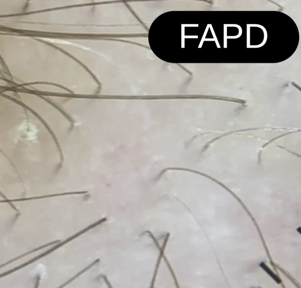
FAPD and Discordant Anisotrichosis — Donovan Hair Clinic
Not every man dressed in a red suit in the month December is Santa Claus. And not every patient with a dual diagnosis of lichen planopilaris (LPP) and androgenetic alopecia (AGA) has fibrosing alopecia in a pattern distribution (FAPD). Over the past few years, I’ve become increasingly concerned (to say it lightly) about a quiet shift in how FAPD is being diagnosed in the world. There is a growing tendency to label patients as having FAPD simply because they carry two diagnoses: lichen planopilaris (LPP) and androgenetic alopecia (AGA). But this is not what FAPD is. So this trend is wrong. FAPD is not merely the coexistence of LPP and AGA. It is a distinct clinicopathologic entity. Patients with FAPD typically show:1) A symmetrical, patterned distribution of hair loss (often central scalp)2)A presentation that mimics AGA—but behaves differently3) Loss of vellus hairs with relative preservation of many isolated terminal hairs4) Subtle but important signs of inflammation, including perifollicular scale or erythema In contrast, classic LPP more often produces patchy, irregular areas of scarring hair loss. The introduction of the concept of discordant anisotrichosis by Saber et al in 2026 has been an important step forward. In typical AGA, hair shaft variability follows a relatively uniform and predictable progression. In FAPD, however, the variability is less than what is expected for the amount of density reduction. This feature provides a practical clue that something more than AGA is occurring. FAPD is not common—and it should not become a default label of AGA+LPP. Cases of FAPD have skyrocketed- and many diagnoses are wrong. The image shown here highlights some key features of FAPD. There is discordant anisotrichosis. Many follicular units contain single terminal hairs. Vellus hairs are not seen. Subtle perifollicular inflammation and scale can also be appreciated. Source link
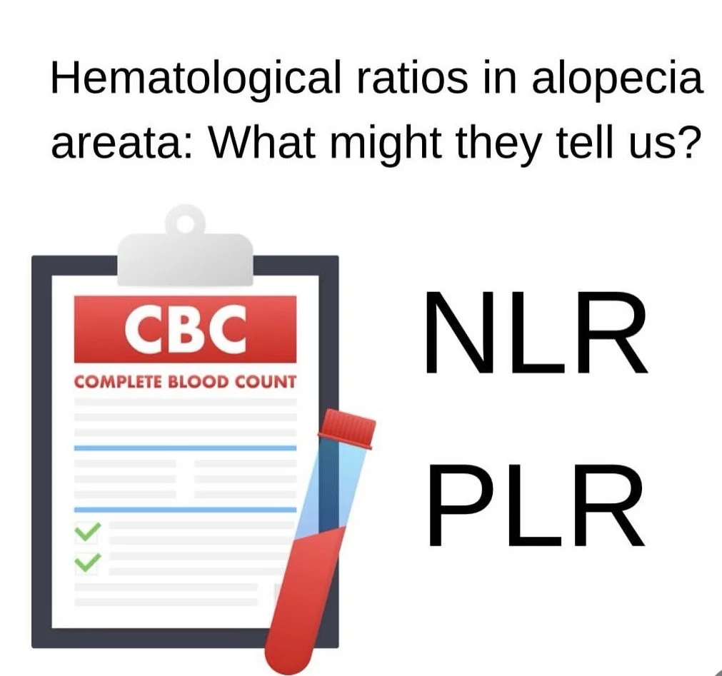
Neutrophil-to-lymphocyte ratios (NLR) and Platelet-to-lymphocyte ratios (PLR) in Alopecia Areata — Donovan Hair Clinic
Every patient with hair loss needs blood tests. Surprisingly, some blood tests reveal more information than you might imagine! For example- a complete blood count (CBC) provides counts of red cells, white cells, and platelets and we can tell if a patient has various issues – like anemia. But there are some interesting information that might also be hidden in test results. Let’s talk about inflammatory indices such as the neutrophil-to-lymphocyte ratio (NLR) and platelet-to-lymphocyte ratio (PLR). Higher values generally reflect a shift toward systemic inflammation. The exact cutoffs and significance is still being studied by researchers but NLR ratios above 3 seems to be concerning and PLR ratios that rise for a given patient over time may also be concerning (although exact cutoff values are less clear) In alopecia areata (AA), several case-control and cross-sectional studies report higher NLR and PLR in patients vs controls. More interestingly, some studies show stepwise increases with disease burden—patients with extensive disease (e.g., higher SALT scores, alopecia totalis/universalis) tend to have higher NLR and PLR than those with patchy AA. A few reports also link higher baseline NLR with longer disease duration and greater activity (e.g., positive hair-pull test), suggesting these ratios may reflect ongoing immune activation. There are also early signals on prognosis and treatment response. Small cohorts have found that lower baseline NLR may be associated with better response to therapies (including corticosteroids and JAK inhibitors), whereas persistently elevated NLR & PLR can track with refractory disease. However, these findings are inconsistent across studies and often lose significance after adjusting for confounders. Importantly, evidence comes almost entirely from small, observational studies with variable cutoffs and methods. There are no validated thresholds and no randomized trials confirming clinical utility. For now, NLR and PLR are best viewed as adjunctive, research-level markers—useful for understanding systemic inflammation in AA, but not reliable standalone tools for diagnosing, staging, or guiding treatment decisions. Source link