
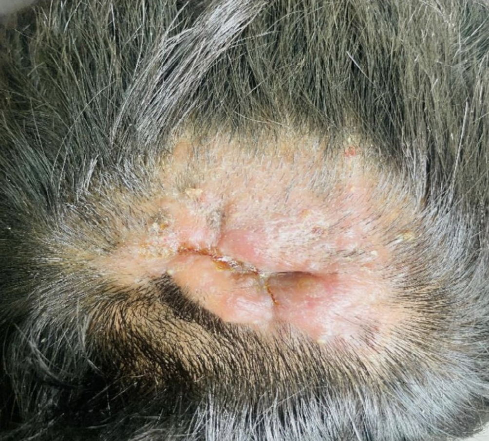
I enjoyed reading a nice paper by Paudyal et al in Clinical Case Reports of a 17 year old male presenting with a scalp abscess that turned out to be a diagnosis of tinea capitis.
Key Lessons from the paper:
1. Kerion can closely mimic a bacterial scalp abscess, leading to unnecessary incision and drainage, antibiotic treatment, and delays in appropriate care.
A painful, swollen, purulent scalp lesion with hair loss should always raise suspicion for kerion (inflammatory tinea capitis). Early recognition can prevent unnecessary surgery and reduce the risk of permanent scarring alopecia.
2. Dermoscopy and a simple KOH examination are invaluable bedside tools for making the diagnosis.
Characteristic dermoscopic findings—including comma hairs, broken hairs of varying lengths, black dots, and perifollicular scaling—combined with a positive KOH preparation can rapidly establish the diagnosis and allow prompt initiation of oral antifungal therapy.
OVERVIEW OF CASE
Kerion is a severe inflammatory form of tinea capitis that can closely resemble bacterial infections such as scalp abscesses or cellulitis. This case report describes a 17-year-old male who presented with a one-month history of a swollen, hairless occipital scalp lesion. He was initially treated by surgeons with incision and drainage followed by two weeks of systemic antibiotics, but failed to improve. After referral to dermatology, dermoscopy revealed classic features of tinea capitis—including comma hairs, black dots, broken hairs, and perifollicular scaling—and a potassium hydroxide (KOH) preparation confirmed dermatophyte infection. The patient was treated with oral griseofulvin, topical clotrimazole, and ketoconazole shampoo, resulting in complete clinical resolution within four weeks and substantial hair regrowth by eight weeks, leaving only minimal residual scarring. This case highlights the importance of considering fungal infection whenever inflammatory scalp lesions are accompanied by alopecia.