

Who Is and Is Not a Candidate This is the most important part of the conversation, and it is why finasteride for women should always involve a thorough consultation rather than a quick prescription. Women who cannot take finasteride include anyone who is pregnant, trying to become pregnant, or not using reliable contraception. Finasteride can cause serious birth defects in male fetuses, and even skin contact with broken or crushed tablets carries a risk. This is a firm contraindication, not a preference. Premenopausal women who are prescribed finasteride off-label must be using reliable birth control and must be counseled clearly on this risk. Women who may be appropriate candidates include postmenopausal women with female pattern hair loss, premenopausal women who are not and do not intend to become pregnant and who are using reliable contraception, and women who have not responded adequately to first-line treatments like minoxidil. Even for appropriate candidates, finasteride is not the right choice for everyone. Hormonal context matters, the underlying cause of the hair loss matters, and whether other factors like thyroid function or iron levels have been addressed first matters. This is why we do not prescribe it in isolation at NHLMA. What Are the Alternatives? Whether finasteride is right for you or not, there are several other treatments with strong evidence for female pattern hair loss worth knowing about. Topical minoxidilis the most established FDA-approved treatment for women with pattern hair loss. It works differently from finasteride, stimulating follicle activity rather than addressing DHT, and it is appropriate for a much broader range of patients including premenopausal women. Low-level laser therapy (LLLT)uses specific wavelengths of light to stimulate follicle metabolism and is a well-tolerated, non-hormonal option that works well as part of a combined protocol. PRP (platelet-rich plasma)and exosome therapy both work by delivering concentrated growth signals directly to the scalp, supporting follicle health and encouraging the transition from resting to active growth phases. These are particularly effective when hair loss is in the earlier stages. Nutritional and hormonal optimization through comprehensive labs is often the missing piece. Addressing iron deficiency, thyroid dysfunction, or hormonal imbalances that are driving the hair loss in the first place can make everything else work better and sometimes resolves shedding on its own. Spironolactone is another off-label option for premenopausal women with androgen-driven hair loss, often considered before finasteride given its longer track record of use in women. What We Actually See in Practice At NHLMA, finasteride is one tool among many, and whether it belongs in a patient’s plan depends on a thorough evaluation first. We look at the type and pattern of hair loss, hormonal and nutritional labs, medical history, and reproductive considerations before discussing it as an option. For postmenopausal women with confirmed androgenetic alopecia who have not responded to first-line treatments, it can be genuinely effective. For younger women, we typically explore other options first and have a detailed conversation about the risks before considering it. What we do not do is treat female hair loss the way male hair loss is treated with a simple prescription and no deeper investigation. The two are fundamentally different problems that require different approaches. Source link

You Have Migraine—Now What? A Step-by-Step Guide To Feeling Better
This post may contain affiliate links. Migraine Strong, as an Amazon Affiliate, makes a small percentage from qualified sales made through affiliate links at no cost to you. Receiving a migraine diagnosis can feel overwhelming. It’s a relief to finally have a name for what you’ve been experiencing, but it also comes with a lot of questions. How do you manage migraine attacks? What migraine treatments are available? What lifestyle changes can make a difference? The good news is that you don’t have to navigate this alone. There are many ways to take control of your health and improve your quality of life. Whether you’re just beginning your journey or looking to refine your management strategies, here are the next steps to consider after a migraine diagnosis. ** While Migraine Strong writes about the latest in migraine treatments, this is not medical advice. We are patient educators. All information you read about should be discussed with your doctor. 1. Find the Right Medical Support: Why a Headache Specialist Matters Migraine is a complex neurological disease, and not all doctors are equipped to provide the specialized care you need. While a general practitioner or general neurologist may have some experience with migraine, a headache specialist has additional training and focuses on treating headache disorders. They can help you develop a personalized treatment plan, identify triggers, and explore cutting-edge therapies. If you have high frequency attacks, headache specialists are your best bet for treatment. How to Find A Headache Specialist Finding a headache specialist can take time, but it’s worth the effort. The American Migraine Foundation and the National Headache Foundation offer directories to help locate specialists. Online support groups can also be a great resource for recommendations. Neura Health is an online neurology clinic. They offer online appointments in a fraction of the time it takes locally. You can see a trained headache provider in a matter of days. They also offer care coaching and a migraine tracker that is reviewed by your headache provider. Read about our experience with Neura Health in this review article. You can also see interviews with various Neura Health providers on the Migraine Strong Instagram. Go to our grid and look for the purple covers indicating what we talked about and with whom. Preparing for Your Appointment To get the most out of your visit, come prepared. Keep a symptom journal detailing your attacks—when they occur, how long they last, and what symptoms accompany them. List past treatments and their effectiveness, and write down any questions you have. The more information you provide, the better your doctor can tailor your treatment. 2. Learn About Available Treatments – Acute vs. Preventive Medications Migraine treatment generally falls into two categories: acute and preventive. Acute medications, such as triptans and gepants, are taken at the onset of an attack to stop it from progressing. Preventive treatments, like beta-blockers, antidepressants, anti-seizure medications, and CGRP inhibitors, aim to reduce the frequency and severity of attacks. CGRP Medications CGRP inhibitors are a breakthrough in migraine treatment. These medications, which include IV infusion (Vyepti), injectables (Aimovig, Ajovy, Emgality) as well as oral options (Qulipta, Nurtec, Ubrelvy, Reyvow, Zavzpret) target a protein involved in migraine attacks. Many patients find relief with these new therapies. Those with commercial insurance can qualify for Patient Assistance programs offered by the pharmaceutical companies which help make the new medications more affordable. Nerve Blocks & Botox For those with frequent attacks, nerve blocks and Botox injections can be effective preventive options. These treatments work by disrupting pain signals and reducing inflammation in the nervous system. Neuromodulation Devices Non-medication options are also available, such as neuromodulation devices like Cefaly, gammaCore, and Nerivio. These devices use electrical pulses to modulate pain pathways and can be a great alternative for those who prefer non-pharmaceutical approaches. 3. Consider Supplements That May Help Many migraine specialists recommend supplements as part of a comprehensive treatment plan. Some of the most commonly suggested options include: Magnesium glycinate, threonate or malate: Can help with brain excitability and reduce attack frequency. These are also better tolerated than magnesium oxide which is the most studied, but often causes intestinal issues. Riboflavin (B2): May improve mitochondrial function and reduce migraine severity. CoQ10: Supports cellular energy production and has been shown to decrease attack frequency. Feverfew & Butterbur: Herbal options that some find beneficial for migraine prevention. Ginger: Helps with nausea and can also treat the symptoms of a migraine attack. When choosing supplements, look for third-party testing to ensure quality and purity. Always consult with your doctor before adding new supplements to your routine. Our Supplement Dispensary offers quality brand supplements and a discount for all of our readers. 4. Prioritize Sleep for Better Migraine Control Why Sleep is Crucial – Sleep disturbances are closely linked to migraine attacks. Poor sleep quality can make attacks more frequent and severe. Tips for Better Sleep Stick to a consistent sleep schedule. Limit screen time before bed. Keep your bedroom cool and dark. Avoid caffeine in the afternoon. If you suspect you have a sleep disorder, such as insomnia or sleep apnea, discuss it with your doctor. Treating underlying sleep issues can significantly improve migraine management. 5. Include Daily Movement in Your Routine Exercise can be a powerful tool for migraine management, but it’s important to approach it carefully. High-intensity workouts and Couch-to-5k programs are best put off until you know how exercise affects your migraine disease. Some people find vigorous exercise triggers migraine attacks. Best Types of Exercise for Migraine Gentle movement is often best, including: Walking Yoga Swimming Low-impact strength training How to Start Without Triggering an Attack Begin slowly and listen to your body. Stay hydrated, warm up properly, and avoid overexertion. If you haven’t exercised in a long time, spend a few weeks ramping up. Start with laps around your living room and gradually expand the amount of time and distance you walk. Over time, regular movement can help reduce attack frequency and improve overall well-being. 6. Discover
#FG Blog- British Society of Gastroenterology minimum service standards and good practice statements for Endoscopic Retrograde Cholangiopancreatography (ERCP)
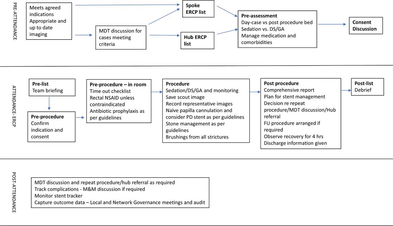
Mohsan Subhani1, Simon M Everett2 Affiliations1School of Medicine University of Nottingham, United Kingdom2Gastroenterology, Leeds Teaching Hospitals NHS Trust, Leeds, UK Endoscopic Retrograde Cholangiopancreatography (ERCP) is one of the highest-risk procedures routinely performed by endoscopists (1). It is one of the most complex endoscopic procedures, often reserved for more advanced therapeutic interventions rather than routine diagnosis. Given its complexity and potential risks, ensuring that the highest standards of care are met is essential. The British Society of Gastroenterology (BSG) first conducted a national audit in 2007 to examine the quality of ERCP training and practice (2). In 2014, BSG published a standard Framework, introducing key performance indicators (KPIs) to guide ERCP practitioners (3). These KPIs focused on practitioner skills, service quality, and training programs to standardise care and improve patient outcomes. ERCP practice has significantly evolved over the years due to the availability of alternative diagnostics tools such as endoscopic ultrasound andmagnetic resonance cholangiopancreatography and it should now mainly be performed for therapeutic purposes. The “Getting it Right First Time” (GIRFT) review and recent NHS white papers highlight the need for multidisciplinary teams, improved case selection, and regional consolidation to enhance quality and outcomes in ERCP (4). In this #FGblog, we want to draw Frontline Gastroenterology readers’ attention to recently published BSG ERCP minimum service standards and good practice statements (5), exploring the driving factors behind these guidelines, and what they mean for both healthcare professionals and patients. In 2021, BSG established a diverse multi-stakeholder ERCP project group including both healthcare professionals and members of the public. Two national surveys were conducted to assess ERCP practices across the UK, targeting both practitioners and endoscopy units. Discussion groups further explored these issues, integrating feedback to draft statements prioritising high-quality, safe ERCP services. The survey had a 100% response rate from all 170 UK endoscopy units and a 74% (389/526 respondents) response from ERCP practitioners. Final recommendations, based on consensus, are presented as good practice statements rather than formal guidelines. These focus on service delivery rather than the technical aspects of performing ERCP. Below is a summary of key statements. The statements are grouped into categories: the patient journey through the unit, the ERCP team, CPD and clinical governance, safety and KPIs, equipment and services, and network provision with multi-disciplinary teams (MDTs). Documents and Policies A high-quality ERCP service requires comprehensive written policies and guidelines. 75% of organisations have clinical pathway guidelines, while only 23% have policies for temporary stent removal. Standard operating procedures (SOPs) reduce variability and enhance safety, as highlighted by reports linking the lack of SOPs to patient safety incidents. ERCP services should have dedicated policies covering vetting, consent, and adherence to published guidelines. Pre-procedure: Referral and Vetting Booking and vetting of ERCP cases should prioritise appropriateness and patient safety. While 58% of units use electronic referral systems, paper-based processes remain common. ERCP consultants are responsible for vetting referrals, ensuring patient fitness and the necessity of procedures. Inpatients require additional review by ERCP team members to avoid late cancellations or inappropriate procedures. Timeliness and Waiting List Management Effective booking and waiting list management are critical for ERCP services. Procedures should be scheduled with proper time allocation to ensure efficiency. Outpatient cases should be prioritized earlier in the day to enable same-day discharge. Additionally, units must track and audit compliance with ERCP timing standards, especially for urgent cases like CBD stones. Monitoring systems for stent removal are crucial to avoid complications such as biliary sepsis. Consent Consent processes for ERCP procedures vary across units, often falling short of BSG and ESGE guidelines. Many patients lack thorough communication about risks, especially in emergency cases. Pre-procedure discussions, preferably face-to-face or by phone, are recommended to ensure informed consent. Final consent must be confirmed by an ERCP-trained endoscopist. Procedure Patients undergoing ERCP often have comorbidities, requiring careful preparation, including medication review, hydration, and imaging. Specialist radiology support is crucial, especially for complex cases. Pre-procedure briefings and adherence to surgical checklists (e.g., WHO) ensure safety, with specific checks for ERCP, such as clotting and stent selection. Managing staff fatigue is important; breaks and lightweight lead gowns should be provided. Teamwork and a learning culture enhance outcomes. Pancreatitis is a common complication; adherence to prevention guidelines and early recognition of complications, like perforation, is vital. Clear escalation pathways should be established to manage adverse events. Post-procedure Recoverypost-procedure care focuses on safe recovery from sedation and monitoring for complications. Patients should be observed for at least four hours after the procedure, with trained nurses available to identify issues like pancreatitis. DischargeDiscussions about the procedure should occur in private after recovery, involving the endoscopist and caregivers. Patients must receive clear discharge information about potential complications and guidance on when to seek help. Follow-upTracking temporary stents is essential to prevent complications. Only 61% of hospitals monitor stent patients, so Trusts must establish reliable systems for timely follow-ups and removals. The ERCP team ERCP nurses must have specific competencies, including leadership skills, to ensure positive patient outcomes. A minimum of three trained staff is essential for each procedure, with one designated as the lead nurse. Management requires coordination between endoscopists, nurses, and administrators to ensure quality and efficiency. Patient engagement in the consent process is critical, as is collaboration with radiologists and surgeons for complex cases. Access to urgent surgical and interventional radiology expertise is essential for safe ERCP procedures, emphasizing the importance of an integrated approach within ERCP networks. Continuous professional development (CPD), clinical governance, safety and KPIs CPD is essential for high-quality patient care, yet many clinicians lack adequate ERCP-related CPD, with 53% having no dedicated time in their job plans. Robust clinical governance is crucial for monitoring safety and quality in high-risk ERCP procedures. KPIs indicate that individual endoscopists should perform a minimum of 100 procedures annually and units should perform a minimum of 200. All ERCP units must implement governance arrangements, designate clinical leads, and ensure compliance with KPIs to improve patient outcomes and care standards. Equipment and services The survey of ERCP facilities highlights areas needing improvement, with

The Weight We Carry: Pain, Resilience, and Recovery in Military Life
By Lexi Mitchell When people think about military service, they often picture discipline, strength, and resilience. What they don’t always see is the hidden cost—the physical and emotional pain that service members carry long after the mission ends. Officer training is infantry-based, and as a smaller female in a physically demanding environment, I learned firsthand how unforgiving that world can be on the body. The Army trains everyone to push beyond limits—but there’s a fine line between endurance and injury. During one combatives training session, I tore my rotator cuff. It was just one of many injuries that came from carrying heavy rucksacks, weapon systems, and gear that easily weighed over 60 pounds—sometimes for miles, over and over again. The physical strain doesn’t discriminate. It doesn’t matter your size or gender—everyone hurts. Feet blister and bleed after a 12-mile ruck march, and yet you wake up in the woods the next day and do it all over again. There’s rarely time for the body to recover before the next test of strength or endurance begins. The culture rewards pushing through pain, and for most of us, stopping wasn’t an option. But pain isn’t just physical. The constant pressure—mental, emotional, and physical—takes a toll that many don’t talk about. Under that much stress, your body’s healing process slows down. Your immune system weakens. When all three—mind, body, and spirit—are under siege, the effects can be long-lasting. Today, I live with the consequences of those years of service. What many people don’t realize is that pain isn’t always visible. I often hear comments like, “You’re young, you look healthy, you look fine.” But the truth is, my pain lies within me. It doesn’t show on my skin, but it affects every part of my life. It’s invisible—but it’s real. I share this not to complain, but to inform. As a veteran and as someone who values facts, I know that stress directly impacts the body’s ability to heal. Chronic pain isn’t weakness—it’s biology. And yet, too many veterans continue to “suck it up,” ignoring pain until it becomes unmanageable. Veterans often carry invisible wounds alongside the visible ones. Pain management isn’t just about medication—it’s about understanding the full human experience of those who served. It’s about listening, validating, and creating care systems that honor both strength and vulnerability. To my fellow veterans: you are not alone. Pain does not define your worth. Seeking help isn’t a sign of weakness—it’s a continuation of your service, because it allows you to reclaim your health and your life. Resources for Veterans in Pain Source link

How Social Prescribing Can Improve Heart Health
Español Heart disease is the number one killer of women in the U.S. Most of us know the standard advice: Exercise more, eat better, quit smoking, manage stress. But even with the best intentions, those instructions can feel abstract, especially if you live in a neighborhood without a safe place to walk, can’t easily afford fresh produce, or go days without talking to another person. What if your healthcare provider could connect you to a community choir, give you a farmers market voucher, or tell you to sign up for a guided walk through your local park and call it medicine? That’s the idea behind social prescribing. What is social prescribing? Social prescribing is a referral system that connects people to non-medical, community-based resources and activities to address the root conditions shaping their health. Think of it as a bridge between your healthcare provider’s office and the rest of life. Julia Hotz, journalist and author of The Connection Cure, the first book written on social prescribing, has followed the movement as it spread from the United Kingdom to more than 32 countries. “Social prescribing got its name from the way it intends to address the social determinants of health,” she explained. “Meaning that people without access to green space, people without access to healthy food and people without access to strong social supports would experience further health disparities.” Hotz identifies five pillars of social prescribing: movement, nature, arts, service and social connection. It’s not a replacement for medication or surgery. But it’s a powerful complement to both, a way of treating the whole person, not just the diagnosis. The hidden heart risks in your social life When most people think of heart disease risk, they think of cholesterol, blood pressure and smoking. But decades of research point to something deeper: Your social world shapes your cardiovascular health in ways that may surprise you. People with poor social health were 30% more likely to develop coronary heart disease and stroke, according to a systematic review of 23 studies. Adults who rarely or never received social support had nearly twice the risk of accumulating three or more cardiovascular risk factors — including hypertension, high cholesterol and diabetes — compared to those with strong support. And loneliness carries its own toll: A 2025 study found that people with chronic loneliness had a 56% higher risk of stroke, even after adjusting for depression and social isolation. Read: How to Make Connections When You’re Lonely >> The biology behind this is straightforward. Stress, loneliness, and depression each elevate cortisol and inflammatory markers, raise blood pressure, and disrupt sleep, all recognized risk factors for heart disease. This is why social prescribing, though it originated as a response to mental health crises, is increasingly recognized as a cardiovascular intervention too. In fact, a 2025 study published in Frontiers in Public Health specifically examined social prescriptions for heart health and found strong relationships between prescriptions involving nature exposure, physical activity and healthy living initiatives and improved cardiovascular outcomes. “It’s true that social prescribing does have a track record of addressing things like stress, loneliness, anxiety, depression, but it can also help heart health in that it could be for activities involving movement, activities involving nature, which are known to de-stress the cardiovascular systems,” Hotz said. Hotz highlights Walk with a Doc, launched by a cardiologist in Columbus, Ohio, and now operating more than 500 chapters worldwide, as a prime example. “I’ve spoken with lots of people who have directly improved their cardiovascular health through these walks,” she said. The power of the prescription Why do patients need a prescription for something they can do on their own? Experts point to the “authority effect” of a formal prescription. When a healthcare provider says a social activity is part of your care plan — not just a nice idea — patients take it more seriously and follow through. “If your doctor says this is an important piece of your health and well-being journey, you’re probably more likely to follow through on that,” said Adrienne Hundley, head of community strategy for SocialRx, a nonprofit that connects patients with arts, cultural and community-based experiences. Having a prescription may also ease the financial burden of participating in community activities by tying them to formal treatment plans so they can be covered by health insurance, which may partner with organizations like SocialRx. Additionally, Medicaid and Medicare Advantage programs increasingly address social determinants of health through wellness initiatives that include social prescription coverage. Beyond insurers, funding can also flow through hospitals’ health equity programs, public health grants, or community nonprofits. SocialRx covers participation costs for members, offers programming in Spanish and other languages, and often includes transportation assistance. Care navigators handle the logistics and serve as accountability partners, checking in after each experience and helping patients stay on track. All of this helps remove barriers that may prevent patients from engaging on their own. SocialRx’s model involves 12 monthly doses of community experience over a year. Hundley shares the story of one older woman who had been nearly housebound for over six months. Her care navigator connected her to a community choir. She went once, came back and by the third month had stepped in as a piano accompanist. By the end, she was the choir’s assistant director at the senior center multiple times a week. “I have found my people, I have found my community, and I have found a renewed lease on life,” she told the team. That kind of transformation can have a powerful physiological impact. According to SocialRx, nearly 4 out of 5 of their members with positive indicators for mental health concerns — anxiety, depression or loneliness — show improvement after their last dose as measured by the The World Health Organization-Five Well-Being Index. Given the links between these conditions and cardiovascular risk, those figures may carry implications for heart health as well. Social prescriptions require a personalized approach Social prescriptions can look wildly different from one person to

Let’s Talk About Vaginismus
Estimated reading time: 5 minutes In the first of our Let’s Talk About series on YouTube, we explore vaginismus. Vaginismus is a form of pelvic floor dysfunction that causes the pelvic floor to involuntarily contract when vaginal penetration is attempted. It can feel as though there is a wall blocking the entrance, which can strike fear into your heart as this is completely outside of your control. With between 5 and 17%1 of women being impacted by the condition, it is critical that we talk about it. In its primary form, vaginismus can prevent penetration when first using tampons or trying to insert a menstrual cup. It can also prevent penetrative sex. For this reason, primary vaginismus is generally discovered during puberty or in early adulthood. The secondary form of vaginismus can occur following childbirth, during menopause or following pelvic surgery or trauma. That trauma can be physical or chemical in nature (such as a bad fall, radiotherapy for cancer treatment or an infection). Lastly, there is a spontaneous form of the condition which can happen at any point in life. This spontaneous is the body’s natural defence mechanism if penetration is forced, but it can also occur when a tampon or vaginal weight is being used. In that instance, the pelvic floor involuntarily contracts preventing removal of the weight or tampon. This is not considered true vaginismus as it’s not a chronic condition and can generally be resolved without extended treatment. How Can You Tell if You Have Vaginismus? Many women mistakenly believe that the initial difficulties, discomfort and pain when first using tampons or trying penetrate sex is vaginismus. It is normal to have some discomfort and pain when you first attempt penetration but it typically resolves over time with practice. In the case of vaginismus, there is no progress even with repeated practice. This is highlighted in the following two graphs. The first shows how discomfort and pain are reduced under normal circumstances as penetration is practiced. Normal Response to Penetration The first shows how discomfort and pain are reduced under normal circumstances as penetration is practiced. Normal pain and discomfort with penetration (when starting out). Vaginismus Response to Penetration The second shows how, with vaginismus, there is no reduction in discomfort or pain, even with persistent practice. Vaginismus pain and discomfort with penetration (when starting out). Studies Into Vaginismus As with other forms of pelvic floor dysfunction, vaginismus isn’t widely studied, however, the studies that have been completed provide some interesting insights into the condition. We have highlighted some studies in this episode. The first revealed heightened sensitivity at rest2 in the pelvic floors of women with vaginismus. If you have read our page on pelvic floor dysfunction, you will have learned that the goal of any pelvic floor training is to return the pelvic floor to an optimal resting length. It is believed3 that the heightened sensitivity experienced by women with vaginismus results in increased pelvic floor hypertonicity. This could be an indication of more fight-or-flight activity in the nervous system. This idea is somewhat bolstered by the second study4 which scanned the brains of women with vaginismus against controls without the condition. That study founds structural changes in the part of the brain responsible for anxiety and fear in the patients with vaginismus, providing the notion that vaginismus may be a fear-related condition. In my book, I explain how the brain cannot tell the difference between what you are thinking and something that is currently happening. Worrying and fearful thoughts have a tendency to place tension into the body, and in the case of vaginismus, this could be a major contributing factor to the condition. Diagnosis and Treatments Getting properly diagnosed and having your vaginismus graded is a first step in your journey towards recovery. In the lower grades, some pelvic floor relaxation, meditation and cognitive behavioural therapy can go a long way towards overcoming the condition. It can be really beneficial to undertake therapy along with your partner as this can help with trust and bonding, which in turn can help communicate a message of safety to your nervous system. In the later grades of vaginismus, dilator therapy may be suggested as an addition to the other exercises and therapies. We’ve shared an earlier article on 5 steps to overcome vaginismus, and have created pages in our knowledge portal sharing information on the condition and it’s conservative and invasive treatments. You will find a lot of information in my book and I have just opened the discounted presale of my course on overcoming vaginismus which will be released on March 18th 2022. If you have questions, jump over to our newly launched community where you will find a space specifically for vaginismus. References Pacik PT, Geletta S. Vaginismus Treatment: Clinical Trials Follow Up 241 Patients. Sex Med. 2017 Jun;5(2):e114-e123. doi: 10.1016/j.esxm.2017.02.002. Epub 2017 Mar 28. PMID: 28363809; PMCID: PMC5440634. Frasson E, Graziottin A, Priori A, Dall’ora E, Didonè G, Garbin EL, Vicentini S, Bertolasi L. Central nervous system abnormalities in vaginismus. Clin Neurophysiol. 2009 Jan;120(1):117-22. doi: 10.1016/j.clinph.2008.10.156. Epub 2008 Dec 13. PMID: 19071060. Unal, Suheyla & Soylu, Ahmet & Yılmaz, Uğur & Karlıdağ, Rifat & Özcan, Abdulcemal & BAydinc, Can. (2002). Vaginismus:Possible role of symphatetic nervous system. 10.13140/RG.2.2.16797.41443. Atmaca M, Baykara S, Ozer O, Korkmaz S, Akaslan U, Yildirim H. Hippocampus and amygdala volumes in patients with vaginismus. World J Psychiatry. 2016 Jun 22;6(2):221-5. doi: 10.5498/wjp.v6.i2.221. PMID: 27354964; PMCID: PMC4919261. Source link
Dissecting Cellulitis and Cardiovascular Risk — Donovan Hair Clinic
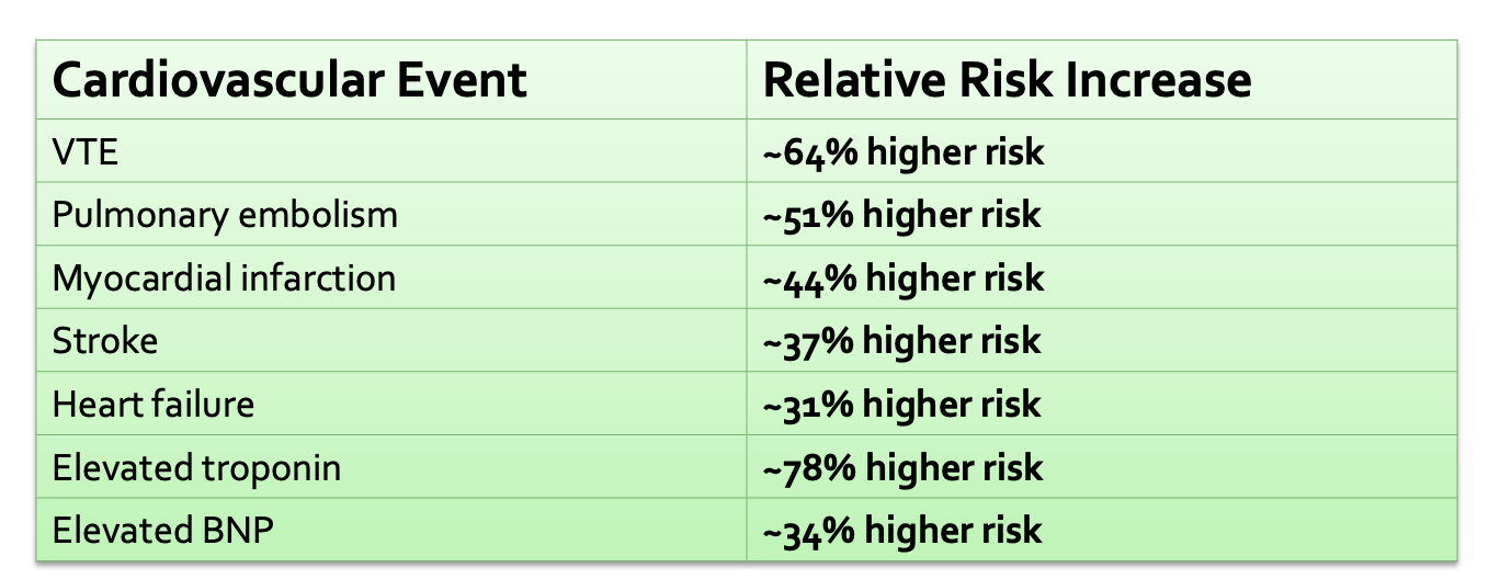
BACKGROUND & PURPOSE Dissecting cellulitis of the scalp (DCS) is a chronic, scarring inflammatory hair disease. It can be painful, disfiguring, and emotionally distressing. Similar inflammatory skin conditions such as hidradenitis suppurativa and psoriasis are linked to higher cardiovascular (CV) disease risk. The purpose of this study was to determine whether patients with DCS experience more cardiovascular problems than similar individuals without DCS. METHODS Researchers used the TriNetX global electronic-health-record database. They identified over 113,000 adults with DCS and matched them 1:1 to individuals without DCS based on age, sex, race, and key medical risk factors. They then compared how often major CV events occurred over time. RESULTS Patients with DCS had significantly higher rates of serious cardiovascular outcomes including myocardial infarction, stroke, pulmonary embolism, venous thromboembolism, atrial fibrillation, and heart failure. VTE showed the largest increased risk—about 64% higher in DCS compared to controls. Source link

Celebrating March for Migraine 2026
2026 was the fifth year of The Migraine Trust’s March for Migraine challenge, where we asked our supporters to walk, run, wheel, hop, skip or even row in one instance, 100 miles across the month of March. A total of 140 people signed up to the challenge and together they raised a fantastic £12,792 which will go towards our work to support people with migraine, fund crucial new research and campaign for change. Not only did our ‘Marchers’ raise vital funds, but they also helped to raise important awareness of migraine and the impact it can have on people’s lives. The majority of our ‘Marchers’ live with migraine themselves, many with chronic migraine, so we are particularly grateful to everyone who took on the challenge while managing migraine attacks or caring for a loved one with migraine. Two of our ‘Marchers’, Bex and Ludmilla gave their reasons as to why they participated: Source link
Mufemilast demonstrates efficacy in ulcerative colitis
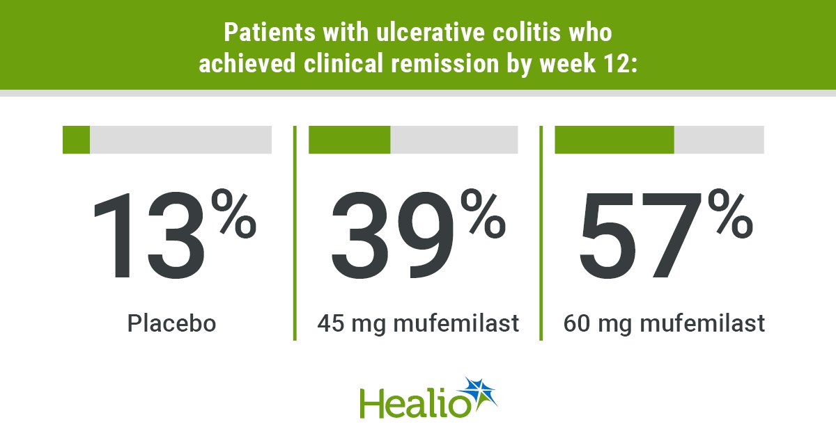
Add topic to email alerts Receive an email when new articles are posted on Please provide your email address to receive an email when new articles are posted on . “ data-action=”subscribe”> Subscribe We were unable to process your request. Please try again later. If you continue to have this issue please contact customerservice@slackinc.com. Back to Healio Key takeaways Mufemilast 60 mg improved clinical remission rate at 12 weeks, which was maintained through 24 weeks. Incidence of adverse events was similar between treatment and placebo groups. CHICAGO — An investigational small-molecule oral phosphodiesterase-4 inhibitor appeared well-tolerated and maintained clinical remission rates at 24 weeks among adults in China with ulcerative colitis, according to data presented. The novel drug, mufemilast (Hemay005, Tianjin Hemay), has been approved in China for patients with plaque psoriasis, but does not have FDA approval. Data derived from Jones CR, et al. Mufemilast (oral pde4 inhibitor) for ulcerative colitis: Open-label extension results at 24 weeks from a multicenter, randomized, double-blind, placebo-controlled parallel group induction phase II clinical trial. Presented at: Digestive Disease Week; May 2-5, 2026; Chicago. Results from a recent trial were presented at Digestive Disease Week. “When looking at this drug, there are some preclinical data that are quite interesting,” presenting author Laurent Peyrin-Biroulet, MD, PhD, head of the IBD unit at Nancy University Hospital in France, told attendees at the meeting. “On top of [an impact on] inflammation, there is also an impact on fibrosis.” Peyrin-Biroulet and colleagues investigated use of mufemilast in UC by conducting a phase 2, double-blind, placebo-controlled trial of 92 patients with moderate to severe disease. Patients needed a modified Mayo score of 4 to 9, an endoscopy score at least 2, and inadequate/failed response or intolerance to conventional therapy or biologics. Participants were randomly assigned to 45 mg mufemilast (n = 31), 60 mg mufemilast (n = 30) or placebo (n = 31) twice daily for 12 weeks, after which all groups switched to 60 mg mufemilast for an additional 12 weeks. Clinical remission rate at week 12 served as the primary endpoint. Secondary endpoints included clinical remission rate at week 24 and clinical response rate at weeks 12 and 24. Analysis included 70 patients who received 60 mg mufemilast in the open treatment period. Results showed clinical remission rates improved from 13% at 12 weeks to 75% at 24 weeks in the group originally assigned placebo, and from 39% to 62% in the group that switched from 45 mg to 60 mg. Remission rates remained steady in the group assigned 60 mg mufemilast for the entire 24-week period (57% to 52%). Researchers also reported increases in clinical response from week 12 to 24 for the groups originally assigned placebo (42% to 81%) and 45 mg mufemilast (87% to 90%), as well as in endoscopic improvement (placebo: 16% to 75%; 45 mg: 42% to 66%). The group assigned 60 mg achieved an 80% clinical response rate at 12 weeks, which dropped slightly to 72% by week 24, and also demonstrated endoscopic improvement (week 12: 67%; week 24: 64%). No significant differences were observed between responses of biologic-naive and biologic-exposed patients. Two adverse events and one severe adverse event — associated with preexisting thrombophlebitis — in the 60 mg group led to trial discontinuation. Incidence of adverse events was comparable between the treatment and placebo groups and included headache, dizziness and nausea. The incidence of headache, which ranged from 9.68% in the placebo and 45 mg groups to 10% in the 60 mg group during the 12-week treatment period, dropped to 2.86% in the open treatment period. Peyrin-Biroulet noted that tuberculosis, which is endemic in China, was an additional safety outcome in the study. “Despite having 17 TB-positive patients receiving mufemilast, no reactivation of TB was observed,” he said. “[Mufemilast] has improved efficacy, significant treatment effect and dose response — which is something that is very important,” Peyrin-Biroulet said. Published by: Sources/Disclosures Source: Jones CR, et al. Mufemilast (oral pde4 inhibitor) for ulcerative colitis: Open-label extension results at 24 weeks from a multicenter, randomized, double-blind, placebo-controlled parallel group induction phase II clinical trial. Presented at: Digestive Disease Week; May 2-5, 2026; Chicago. Disclosures: Healio was unable to confirm relevant financial disclosures at time of publication. Ask a clinical question and tap into Healio AI’s knowledge base. PubMed, enrolling/recruiting trials, guidelines Clinical Guidance, Healio CME, FDA news Healio’s exclusive daily news coverage of clinical data Learn more Add topic to email alerts Receive an email when new articles are posted on Please provide your email address to receive an email when new articles are posted on . “ data-action=”subscribe”> Subscribe We were unable to process your request. Please try again later. If you continue to have this issue please contact customerservice@slackinc.com. Back to Healio Source link
New EDS Patient: Where Do I Begin?
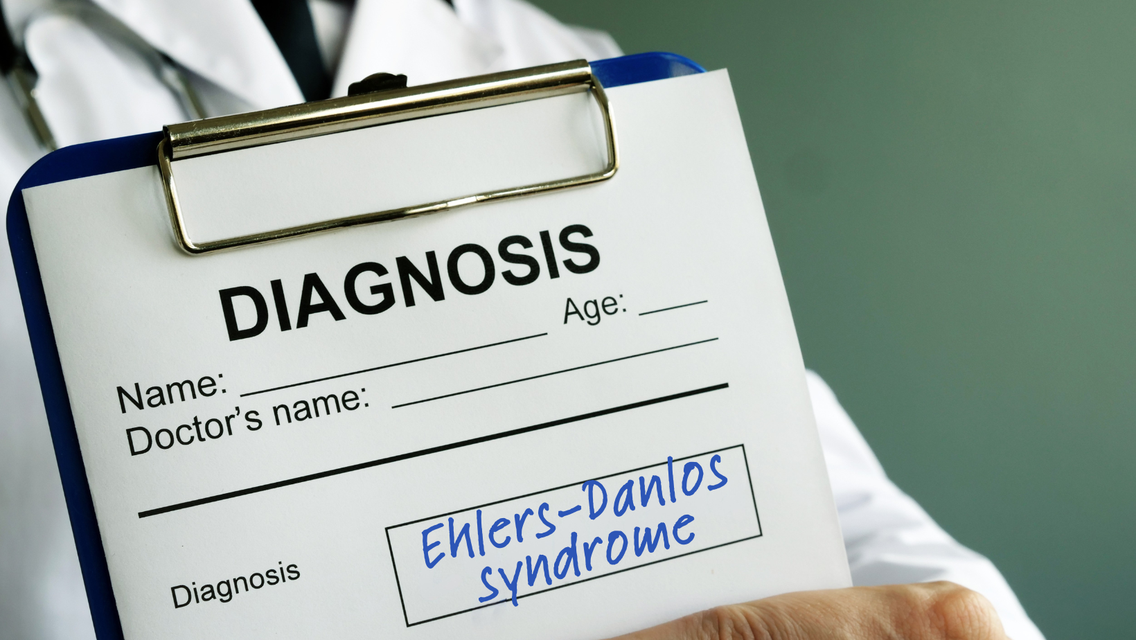
Did you recently get diagnosed with Ehlers-Danlos syndrome (EDS), and feel overwhelmed and confused about where to turn for help? What was described to me years ago as a rare condition is now believed to be much more prevalent, particularly when combined with the broader diagnosis of joint hypermobility disorder or hypermobility spectrum disorder (HSD). Still, many, like me, wait years to find out what is really the problem. Being diagnosed at the age of 54 with a condition I was born with lit a spark in me to do all I could to help others learn to cope. We have to wonder, if so many of us are living with the chaos and confusion of this condition, then why do we feel so lost as to where to begin to help ourselves? Shouldn’t the medical field be right there by our side with guidance? Well, as they catch up to us and learn how to help us, let’s make it our mission to pass information forward to help those around us. That may look like setting up a support group, sharing a list of medical personnel who are willing to learn and help with EDS, and paying it forward by sharing the strategies that have worked for us—including to medical providers working to learn more, as I’ve done in the past. But meanwhile, if you’ve just been diagnosed or don’t know where to start, what can you do to begin the process of trying to get on top of this condition? This Ehlers-Danlos Syndromes (EDS) and Hypermobility Spectrum Disorders (HSD) Awareness Month, I’d like to share some of what I’ve experienced and learned during my decades of living with and managing this disease. Please note that the information in this article should not be considered as professional medical advice, diagnosis, or treatment. It is for informational purposes only and represents my opinions alone, rather than the views of the U.S. Pain Foundation. These are my personal experiences and suggestions on what has helped me improve my quality of life. I share this in hopes that others living with this condition might also learn something new and improve their quality of life, too. What is Ehlers-Danlos syndrome? EDS is an inherited disorder primarily involving problems with the body’s connective tissue. There are 13 subtypes, with various levels of severity and impact. Hypermobile EDS (hEDS) is by far the most common subtype, accounting for about 9 in 10 cases. Classical EDS (cEDS) and vascular EDS (vEDS) are the next most common types, with the remaining subtypes being diagnosed much more rarely. HSD more broadly describes connective tissue disorders causing joint hypermobility and instability; some people are diagnosed with HSD when they don’t meet all of the diagnostic criteria for EDS. While this piece is largely based on my experiences with EDS, many of these insights will apply to those with HSD as well. Some people living with EDS go through life with minimal symptoms and little pain. Others are faced with a wide range of symptoms and complications. Clinical manifestations of EDS are most often joint- and skin-related and may include: Joints: Joint hypermobility (extreme flexibility) and hyperextensibility (movement beyond the joint’s normal range); loose or unstable joints that are prone to frequent dislocations and/or subluxations; joint pain. Skin: Soft, velvety skin; skin hyperextensibility (stretchiness); fragile skin that tears or bruises easily or severely; severe scarring; slow and poor wound healing. Other symptoms, which can vary by subtype, include chronic, early-onset, debilitating musculoskeletal pain; fatigue; scoliosis or neck instability; and mitral valve prolapse. Less common symptoms or complications associated with rare subtypes can include arterial, intestinal, or uterine fragility or rupture; scleral fragility; poor muscle tone; and gum disease. Due in part to the fact that the connective tissue impacted by EDS is widespread throughout the body, there are a number of associated disorders people may deal with, including postural orthostatic tachycardia syndrome (POTS) and other types of dysautonomia; mast cell activation syndrome (MCAS); Raynaud’s disease; early-onset osteoarthritis; autoimmune conditions; migraine and other headache disorders; a number of gastrointestinal disorders; gynecologic conditions; Chiari I malformation; craniocervical instability; tethered cord syndrome; and more. EDS is often misdiagnosed as fibromyalgia, osteoarthritis, rheumatoid arthritis, lupus, multiple sclerosis, myalgic encephalomyelitis/chronic fatigue syndrome (ME/CFS), or Munchausen syndrome or other psychiatric disorders. To complicate matters further, it is possible to have both EDS alongside one or more of most of these disorders. How is EDS Diagnosed? While a number of providers or specialists, including general practitioners or rheumatologists, may be initially involved in identifying or suspecting EDS, a confirmed diagnosis is often obtained by a geneticist familiar with the disease, as our symptoms can vary significantly. For some types of EDS, a skin biopsy to determine the chemical makeup of the connective tissue can help to suggest the diagnosis. Genetic testing can also help identify most subtypes of EDS—but not hEDS, the most common type. Geneticists often diagnose hEDS after ruling out other types. For some, the diagnosis of EDS is based upon the patient’s clinical findings and family history. However, while family history is key in helping diagnose EDS, particularly when genetic testing can’t help, the National Institutes of Health (NIH) has found patients who were born with EDS with no family history. And due to the difficulty of receiving a diagnosis, some people are the first in their family to be diagnosed. After receiving a diagnosis of EDS, you can work with your providers to find the best ways to manage the disease. These are some of the tips I have learned. Maintaining Physical Activity When you live with chronic pain, you can get emotionally and physically worn down, and you may sometimes feel that you have no energy to exercise. But living with EDS makes it all the more important to do just that. We need to keep our muscles strong to help support our joints, and cardio workouts also help keep our bodies in the best shape possible. Consider the stationary bike,